. 2011 Sep;193(18):4904-13.
doi: 10.1128/JB.05231-11. Epub 2011 Jul 15.

Roles of the redox-active disulfide and histidine residues forming a catalytic dyad in reactions catalyzed by 2-ketopropyl coenzyme M oxidoreductase/carboxylase

Affiliations

Roles of the redox-active disulfide and histidine residues forming a catalytic dyad in reactions catalyzed by 2-ketopropyl coenzyme M oxidoreductase/carboxylase

Melissa A Kofoed et al. J Bacteriol. 2011 Sep.

Abstract

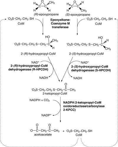
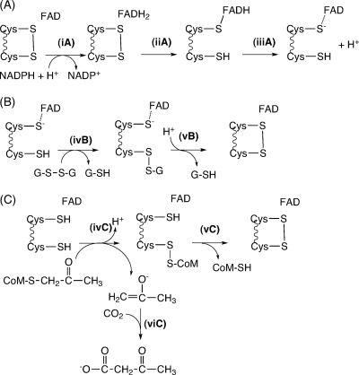
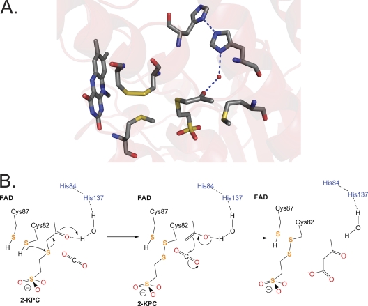
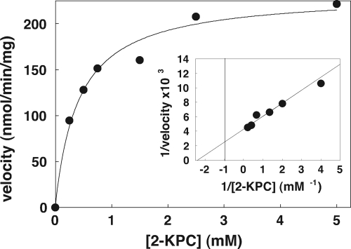
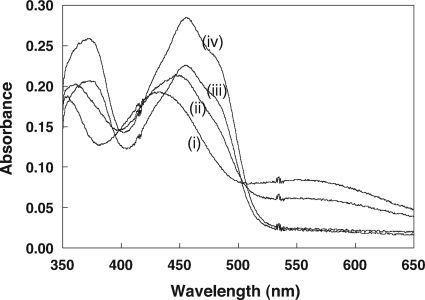
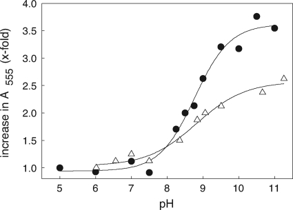
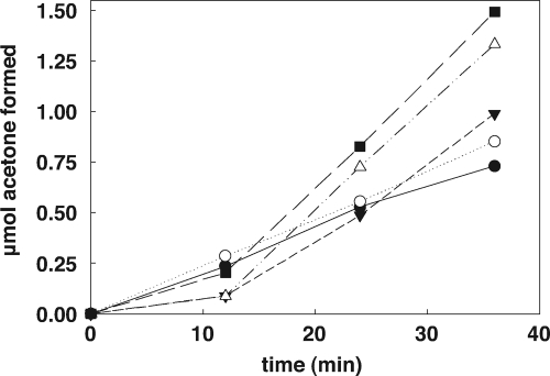
NADPH:2-ketopropyl-coenzyme M oxidoreductase/carboxylase (2-KPCC), an atypical member of the disulfide oxidoreductase (DSOR) family of enzymes, catalyzes the reductive cleavage and carboxylation of 2-ketopropyl-coenzyme M [2-(2-ketopropylthio)ethanesulfonate; 2-KPC] to form acetoacetate and coenzyme M (CoM) in the bacterial pathway of propylene metabolism. Structural studies of 2-KPCC from Xanthobacter autotrophicus strain Py2 have revealed a distinctive active-site architecture that includes a putative catalytic triad consisting of two histidine residues that are hydrogen bonded to an ordered water molecule proposed to stabilize enolacetone formed from dithiol-mediated 2-KPC thioether bond cleavage. Site-directed mutants of 2-KPCC were constructed to test the tenets of the mechanism proposed from studies of the native enzyme. Mutagenesis of the interchange thiol of 2-KPCC (C82A) abolished all redox-dependent reactions of 2-KPCC (2-KPC carboxylation or protonation). The air-oxidized C82A mutant, as well as wild-type 2-KPCC, exhibited the characteristic charge transfer absorbance seen in site-directed variants of other DSOR enzymes but with a pK(a) value for C87 (8.8) four units higher (i.e., four orders of magnitude less acidic) than that for the flavin thiol of canonical DSOR enzymes. The same higher pK(a) value was observed in native 2-KPCC when the interchange thiol was alkylated by the CoM analog 2-bromoethanesulfonate. Mutagenesis of the flavin thiol (C87A) also resulted in an inactive enzyme for steady-state redox-dependent reactions, but this variant catalyzed a single-turnover reaction producing a 0.8:1 ratio of product to enzyme. Mutagenesis of the histidine proximal to the ordered water (H137A) led to nearly complete loss of redox-dependent 2-KPCC reactions, while mutagenesis of the distal histidine (H84A) reduced these activities by 58 to 76%. A redox-independent reaction of 2-KPCC (acetoacetate decarboxylation) was not decreased for any of the aforementioned site-directed mutants. We interpreted and rationalized these results in terms of a mechanism of catalysis for 2-KPCC employing a unique hydrophobic active-site architecture promoting thioether bond cleavage and enolacetone formation not seen for other DSOR enzymes.


Figures

Fig. 1.
Fig. 1.
Pathway of aliphatic epoxide carboxylation in Xanthobacter autotrophicus Py2 and Rhodococcus rhodochrous B276.
Fig. 2.
Fig. 2.
Reactions of members of the DSOR family. (A) Steps resulting in the reduction of the redox-active cysteine disulfide for all DSOR enzymes; (B) steps resulting in reduction of a substrate with an oxidized disulfide bond, as illustrated for glutathione reductase; (C) steps resulting in reduction of the thioether bond of 2-KPC in 2-KPCC, resulting in the production of enolacetone, which undergoes carboxylation to form acetoacetate. Note that the reduced flavin cysteine is shown in the thiol rather than as the thiolate form for 2-KPCC for reasons described in the paper.
Fig. 3.
Fig. 3.
Active-site architecture and proposed mechanism for 2-KPCC. (A) Structure of 2-KPC bound to 2-KPCC, highlighting active-site residues believed to be key to catalysis (Protein Data Bank ID, 1MO9). (B) Proposed mechanism of thioether bond cleavage, enolacetone formation and stabilization, and carboxylation based on the structures solved for 2-KPCC and the results of the present work. The initial abstraction of a proton from C82 may be facilitated by a general base that has not yet been identified. The reduction of the mixed disulfide of CoM and Cys82 is not shown but will occur as for glutathione reductase (Fig. 2B).
Fig. 4.
Fig. 4.
SDS-PAGE and Western blotting of recombinant 2-KPCC. (A) SDS-polyacrylamide gel. Lanes: 1, molecular mass standards; 2, cell extract (13.3 μg); 3, first Ni2+ fraction (6.7 μg); 4, phenyl-Sepharose fraction (6.2 μg); 5, enterokinase digestion (15 μg); 6, the eluate from the second Ni2+ affinity column (15 μg). (B) Immunoblot-prepared blot from an identical SDS-polyacrylamide gel using antibodies raised to native 2-KPCC.
Fig. 5.
Fig. 5.
Effect of 2-KPC concentration on the reductive cleavage and carboxylation of 2-KPC to acetoacetate and CoM. All assays were performed with 0.089 mg of 2-KPCC. The line through the data points was generated by nonlinear least-squares fitting to a rectangular hyperbola. Inset, double-reciprocal plot of the data. The line through the data points was also generated by nonlinear least-squares fitting of the data.
Fig. 6.
Fig. 6.
UV/visible absorption spectra of the r-2-KPCC C82A mutant at various pH values. Spectra were obtained using 0.15 mg of 2-KPCC diluted to a volume of 130 μl as described in Materials and Methods. The spectral noise at ∼A420 and ∼A535 is due to the ultramicro-sized cuvette that was used. The spectra shown are for pH values of (i) 11.0, (ii) 9.0, (iii) 7.5, and (iv) 5.0.
Fig. 7.
Fig. 7.
Determination of pKa values for the interchange thiol of 2-KPCC. The fold increase in A555 due to formation of charge transfer absorbance between FAD and the thiolate of C87 is plotted versus pH. The lines were derived from the four-parameter sigmoidal curve fit used to calculate pKa values. Symbols: •, C82A mutant of r-2-KPCC; ▵, native 2-KPCC alkylated on C82 with BES.
Fig. 8.
Fig. 8.
Acetoacetate decarboxylase activity of wild-type and mutant r-2-KPCC proteins. Assays contained 0.125 mg of 2-KPC, 250 mM acetoacetate, and 5 mM CoM. Data points represent the averages of duplicate experiments. Symbols: •, wild-type r-2-KPCC; ○, H84A r-2-KPCC; ▾, H137A r-2-KPCC; ▵, C87A r-2-KPCC; ▪, C82A r-2-KPCC.

References

    1. Allen J. R., Clark D. D., Krum J. G., Ensign S. A. 1999. A role for coenzyme M (2-mercaptoethansulfonic acid) in a bacterial pathway of aliphatic epoxide carboxylation. Proc. Natl. Acad. Sci. U. S. A. 96:8432–8437 - PMC - PubMed
    1. Allen J. R., Ensign S. A. 1996. Carboxylation of epoxides to β-keto acids in cell extracts of Xanthobacter strain Py2. J. Bacteriol. 178:1469–1472 - PMC - PubMed
    1. Allen J. R., Ensign S. A. 1997. Characterization of three protein components required for functional reconstitution of the epoxide carboxylase multienzyme complex from Xanthobacter strain Py2. J. Bacteriol. 179:3110–3115 - PMC - PubMed
    1. Allen J. R., Ensign S. A. 1998. Identification and characterization of epoxide carboxylase activity in cell extracts of Nocardia corallina strain B276. J. Bacteriol. 180:2072–2078 - PMC - PubMed
    1. Allen J. R., Ensign S. A. 1997. Purification to homogeneity and reconstitution of the individual components of the epoxide carboxylase multiprotein enzyme complex from Xanthobacter strain Py2. J. Biol. Chem. 272:32121–32128 - PubMed

Publication types

LinkOut - more resources