

doi: 10.1128/JB.05485-11.
Epub 2011 Jul 15.
Characterization of components of the Staphylococcus aureus mRNA degradosome holoenzyme-like complex
Affiliations
- PMID: 21764917
- PMCID: PMC3187390
- DOI: 10.1128/JB.05485-11
Item in Clipboard
Characterization of components of the Staphylococcus aureus mRNA degradosome holoenzyme-like complex
J Bacteriol.
2011 Oct.
Abstract
Bacterial two-hybrid analysis identified the Staphylococcus aureus RNA degradosome-like complex to include RNase J1, RNase J2, RNase Y, polynucleotide phosphorylase (PNPase), enolase, phosphofructokinase, and a DEAD box RNA helicase. Results also revealed that the recently recognized RNase RnpA interacts with the S. aureus degradosome and that this interaction is conserved in other Gram-positive organisms.
Figures
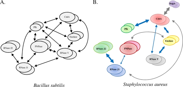
B. subtilis and S. aureus mRNA degradosome-like complexes. (A) B. subtilis RNA degradosome-like complex (6). (B) Deduced S. aureus RNA degradosome-like complex. Blue arrows represent conserved interactions between S. aureus and B. subtilis proteins. Arrow thickness indicates the strength of interactions as determined by quantitative β-galactosidase activity results (see Table S2 in the supplemental material).
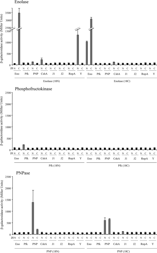
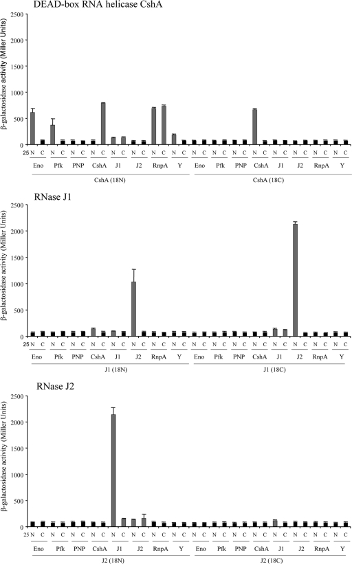
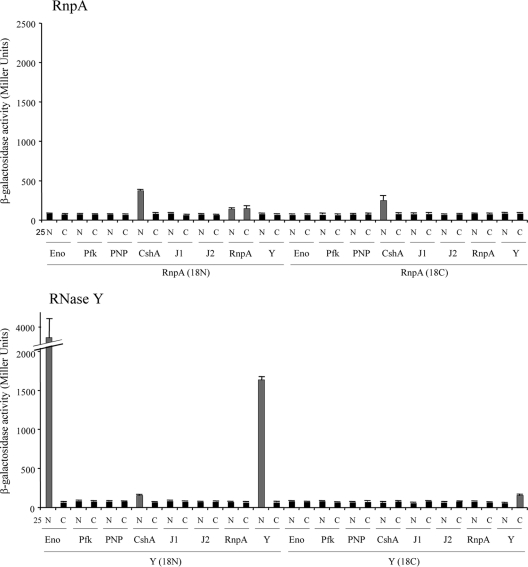
β-Galactosidase-deduced protein-protein interactions. β-Galactosidase activities of transformants with the indicated S. aureus proteins fused to the N terminus (N) or C terminus (C) of the T25 and T18 catalytic fragments of CyaA. Gray bars represent statistically significant, positive interactions of proteins/domains, whereas black bars represent negative interactions. S. aureus proteins tested included enolase (Eno), phosphofructokinase (Pfk), PNPase (PNP), DEAD box RNA helicase CshA, RNase J1 (J1), RNase J2 (J2), RnpA, and RNase Y (Y).
β-Galactosidase-deduced protein-protein interactions. β-Galactosidase activities of transformants with the indicated S. aureus proteins fused to the N terminus (N) or C terminus (C) of the T25 and T18 catalytic fragments of CyaA. Gray bars represent statistically significant, positive interactions of proteins/domains, whereas black bars represent negative interactions. S. aureus proteins tested included enolase (Eno), phosphofructokinase (Pfk), PNPase (PNP), DEAD box RNA helicase CshA, RNase J1 (J1), RNase J2 (J2), RnpA, and RNase Y (Y).
β-Galactosidase-deduced protein-protein interactions. β-Galactosidase activities of transformants with the indicated S. aureus proteins fused to the N terminus (N) or C terminus (C) of the T25 and T18 catalytic fragments of CyaA. Gray bars represent statistically significant, positive interactions of proteins/domains, whereas black bars represent negative interactions. S. aureus proteins tested included enolase (Eno), phosphofructokinase (Pfk), PNPase (PNP), DEAD box RNA helicase CshA, RNase J1 (J1), RNase J2 (J2), RnpA, and RNase Y (Y).
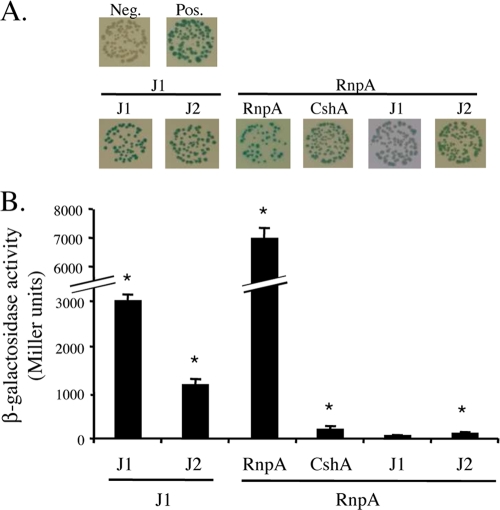
Bacterial two-hybrid analyses of selected B. subtilis proteins. (A) Qualitative analysis of the β-galactosidase activities of the indicated protein pairs. (B) Corresponding quantitative analysis of β-galactosidase activity. * indicates a P value of ≤0.01 (see Table S4 in the supplemental material). B. subtilis proteins tested were RNase J1 (J1), RNase J2 (J2), CshA, and RnpA.
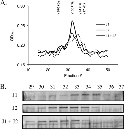
In vitro interaction of purified S. aureus RNase J1 and RNase J2. (A) Gel filtration analysis of RNase J1, RNase J2, and RNase J1 plus RNase J2, as measured by determining the optical density at 595 nm (OD595). Molecular mass size standards are shown above the chromatogram. (B) Silver-stained 10% SDS-PAGE of eluted RNase J1, RNase J2, and RNase J1 plus RNase J2. Elution fractions are indicated above the gels.
References
-
- Carpousis A. J. 2002. The Escherichia coli RNA degradosome: structure, function and relationship in other ribonucleolytic multienzyme complexes. Biochem. Soc. Trans. 30:150–155 - PubMed
-
- Carpousis A. J., Van Houwe G., Ehretsmann C., Krisch H. M. 1994. Copurification of E. coli RNase E and PNPase: evidence for a specific association between two enzymes important in RNA processing and degradation. Cell 76:889–900 - PubMed
-
- Chandran V., Luisi B. F. 2006. Recognition of enolase in the Escherichia coli RNA degradosome. J. Mol. Biol. 358:8–15 - PubMed
Publication types
MeSH terms
Substances
Grants and funding
LinkOut - more resources
Full Text Sources
Other Literature Sources
Molecular Biology Databases
