. 2011 Sep;193(18):4685-98.
doi: 10.1128/JB.05483-11. Epub 2011 Jul 15.

Systematic analysis of diguanylate cyclases that promote biofilm formation by Pseudomonas fluorescens Pf0-1

Affiliations

Systematic analysis of diguanylate cyclases that promote biofilm formation by Pseudomonas fluorescens Pf0-1

Peter D Newell et al. J Bacteriol. 2011 Sep.

Abstract

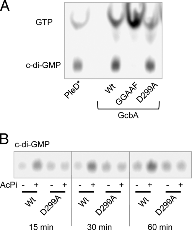
Cyclic di-GMP (c-di-GMP) is a broadly conserved, intracellular second-messenger molecule that regulates biofilm formation by many bacteria. The synthesis of c-di-GMP is catalyzed by diguanylate cyclases (DGCs) containing the GGDEF domain, while its degradation is achieved through the phosphodiesterase activities of EAL and HD-GYP domains. c-di-GMP controls biofilm formation by Pseudomonas fluorescens Pf0-1 by promoting the cell surface localization of a large adhesive protein, LapA. LapA localization is regulated posttranslationally by a c-di-GMP effector system consisting of LapD and LapG, which senses cytoplasmic c-di-GMP and modifies the LapA protein in the outer membrane. Despite the apparent requirement for c-di-GMP for biofilm formation by P. fluorescens Pf0-1, no DGCs from this strain have been characterized to date. In this study, we undertook a systematic mutagenesis of 30 predicted DGCs and found that mutations in just 4 cause reductions in biofilm formation by P. fluorescens Pf0-1 under the conditions tested. These DGCs were characterized genetically and biochemically to corroborate the hypothesis that they function to produce c-di-GMP in vivo. The effects of DGC gene mutations on phenotypes associated with biofilm formation were analyzed. One DGC preferentially affects LapA localization, another DGC mainly controls swimming motility, while a third DGC affects both LapA and motility. Our data support the conclusion that different c-di-GMP-regulated outputs can be specifically controlled by distinct DGCs.


Figures

Fig. 1.
Fig. 1.
Genetic characterization of gcbA. (A) A quantitative biofilm assay comparing the WT, Pfl01_0623 transposon mutant, and unmarked gcbA deletion strains. Data presented are the mean absorbances of dissolved crystal violet-stained biofilm material ± standard deviations (SD) (n = 12). Representative images are shown above the graph. (B) A schematic of the GcbA protein showing the amino acid numbers and predicted domains. The lowercase “rec” indicates a degenerate REC domain. (C) A quantitative biofilm assay examining the complementation of the ΔgcbA mutant biofilm phenotype. pMQ72 is the empty complementation vector; 0.2% arabinose was added to the medium. (D) A representative image of a swimming motility assay in 0.35% agar assessing the complementation of the ΔgcbA mutant. (E) Quantitative measurements of swim zone areas are presented for eight replicates of the assay shown in panel D. Data presented are mean percentages of WT swim areas on the same plate ± standard errors (SE) (n = 8).
Fig. 2.
Fig. 2.
Enzymatic characterization of GcbA. (A) In vitro DGC assays comparing GcbA and mutant variants to the control DGC, PleD*, are shown resolved on a thin-layer chromatography plate. Compounds are labeled on the left; the image was generated by a phosphor screen scanner. (B) Assay comparing the effect of acetylphosphate (AcPi) treatment on the DGC activity of WT GcbA and the D299A mutant.
Fig. 3.
Fig. 3.
Effect of mutations in putative DGCs on biofilm formation. (A) A quantitative biofilm assay analyzing the WT and single-crossover insertion mutants in gcbA and 29 putative DGCs; data are mean percentages of the WT ± SD (n = 8). Asterisks indicate a statistically significant decrease in biofilm relative to that of the WT (P < 0.01 in a two-tailed Student's t test assuming equal variance). (B) A quantitative biofilm assay comparing the WT and strains with deletion mutations of Pfl01_0623 (ΔgcbA), Pfl01_1789 (ΔgcbB), Pfl01_4666 (ΔgcbC), and Pfl01_1058 (ΔwspR).
Fig. 4.
Fig. 4.
Complementation analysis of wspR. (A) A quantitative biofilm assay examining the complementation of the ΔwspR mutant phenotype; 0.1% arabinose was added to the medium. (B) Western blot comparing the abundance of WspR-6H and WspR-GGAAF proteins in vivo. Samples were probed for the 6×His epitope on each protein, and cells were cultured in K10T-1 with 0.1% arabinose.
Fig. 5.
Fig. 5.
Complementation analysis of gcbB. (A) A schematic of the GcbB protein showing the amino acid numbers (above) and predicted domains in boxes. TM, transmembrane. (B) Western blot comparing the abundance of GcbB-6H and GcbB-GGAAF proteins in vivo. Samples were probed for the 6×His epitope on each protein, and cells were cultured in K10T-1 with 0.01% arabinose. (C) A quantitative biofilm assay examining the complementation of the ΔgcbB mutant phenotype by pGcbB-6H and pGcbB-GGAAF at three concentrations of inducer (ara, arabinose). (D) A quantitative immunoblot assay probing whole cells for the surface expression of LapA. Pixel densities of blot images were calculated using ImageJ, and the mean density for the WT was set to 100. Data shown are means ± SE (n = 7). Representative images are shown above the graph.
Fig. 6.
Fig. 6.
Complementation analysis of gcbC. (A) A schematic of the GcbC protein showing the amino acid numbers and predicted domains in boxes. TM, transmembrane. (B) Western blot comparing the abundance of GcbC-HA and GcbC-GGAAF proteins in vivo. Samples were probed for HA on each protein, and cells cultured in K10T-1 without arabinose. (C) A quantitative biofilm assay examining the complementation of the ΔgcbC mutant phenotype by pGcbC-HA and pGcbC-GGAAF with and without inducer (ara, arabinose). (D) A quantitative immunoblot assay probing whole cells for the surface expression of LapA. Data shown are means ± SE (n = 7). Representative images are shown above the graph. (E) A representative image of a swimming motility assay in 0.35% agar, assessing the complementation of the ΔgcbC mutant. (F) Quantitative measurements of swim zone areas are presented for eight replicates of the assay shown in panel E. Data presented are mean percentages of WT swim area on the same plate ± SE (n = 8).
Fig. 7.
Fig. 7.
In vitro and in vivo assessments of DGC activity. (A) In vitro DGC assays comparing GcbC-Hyb and GcbC-Hyb-GGAAF mutant variants to GcbA as a positive control. An image of a thin-layer chromatography plate generated by a phosphor screen scanner is shown with compounds labeled on the left. (B) Quantitative measurements of cellular c-di-GMP from the Δ4DGC strain expressing the indicated plasmids. Formic acid-extracted c-di-GMP was measured by LC/MS and normalized to mg of dry weight of bacteria after extraction.
Fig. 8.
Fig. 8.
Combinatorial analysis of DGC mutants. (A) A quantitative biofilm assay comparing each single-, double-, triple-, and quadruple-mutant combination for the DGC genes indicated (means ± SD; n = 8). (B) A quantitative immunoblot assay probing whole cells for the surface expression of LapA by the indicated strains. Data shown are means ± SE (n = 7). Representative images are shown above the graph. (C) Quantitative swimming motility assay for the strains indicated. Data presented are mean percentages of WT swim area on the same plate ± SE (n = 8). Representative images of each mutant and adjacent WT control are shown above the corresponding bar in the graph.

References

    1. An S., Wu J., Zhang L. H. 2010. Modulation of Pseudomonas aeruginosa biofilm dispersal by a cyclic-Di-GMP phosphodiesterase with a putative hypoxia-sensing domain. Appl. Environ. Microbiol. 76:8160–8173 - PMC - PubMed
    1. Banin E., Vasil M. L., Greenberg E. P. 2005. Iron and Pseudomonas aeruginosa biofilm formation. Proc. Natl. Acad. Sci. U. S. A. 102:11076–11081 - PMC - PubMed
    1. Bantinaki E., et al. 2007. Adaptive divergence in experimental populations of Pseudomonas fluorescens. III. Mutational origins of wrinkly spreader diversity. Genetics 176:441–453 - PMC - PubMed
    1. Choy W. K., Zhou L., Syn C. K., Zhang L. H., Swarup S. 2004. MorA defines a new class of regulators affecting flagellar development and biofilm formation in diverse Pseudomonas species. J. Bacteriol. 186:7221–7228 - PMC - PubMed
    1. Christen M., Christen B., Folcher M., Schauerte A., Jenal U. 2005. Identification and characterization of a cyclic di-GMP-specific phosphodiesterase and its allosteric control by GTP. J. Biol. Chem. 280:30829–30837 - PubMed

Publication types

MeSH terms