. 2011 Sep;193(18):4841-8.
doi: 10.1128/JB.05258-11. Epub 2011 Jul 15.

Bacillus subtilis CodY operators contain overlapping CodY binding sites

Affiliations

Bacillus subtilis CodY operators contain overlapping CodY binding sites

Lewis V Wray Jr et al. J Bacteriol. 2011 Sep.

Abstract

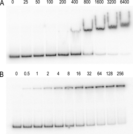
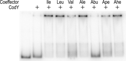
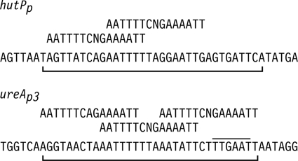
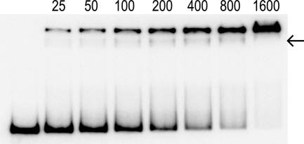
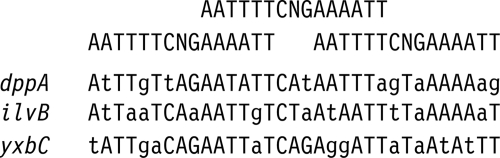
CodY is a global transcriptional regulator that is activated by branched-chain amino acids. A palindromic 15-bp sequence motif, AATTTTCNGAAAATT, is associated with CodY DNA binding. A gel mobility shift assay was used to examine the effect of pH on the binding of Bacillus subtilis CodY to the hutPp and ureAp(3) promoters. CodY at pH 6.0 has higher affinity for DNA, more enhanced activation by isoleucine, and a lower propensity for nonspecific DNA binding than CodY at pH 8.0. DNase I footprinting was used to identify the CodY-protected regions in the hutPp and ureAp(3) promoters. The CodY-protected sequences for both promoters were found to contain multiple copies of the 15-bp motif with 6-bp overlaps. Mutational analysis of the hutPp regulatory region revealed that two overlapping sequence motifs were required for CodY-mediated regulation. The presence of overlapping sequence motifs in the regulatory regions of many B. subtilis CodY-regulated genes suggests that CodY binds to native operators that contain overlapping binding sites.


Figures

Fig. 1.
Fig. 1.
CodY-hutPp gel mobility shift assays. These assays were performed at pH 8.0 (A) and at pH 6.0 (B). Isoleucine (32 mM) was included in the binding reaction mixture, gel, and gel buffer. The numbers above each lane correspond to the CodY dimer concentration (nM).
Fig. 2.
Fig. 2.
Enhancement of CodY DNA-binding activity by aliphatic amino acids. The experiments were performed at pH 6.0. CodY with a dimer concentration of 200 nM was present where indicated. All of the amino acids were l-stereoisomers and were present only in the binding reaction sample. Nonstandard abbreviations: Ale, alloisoleucine; Abu, 2-aminobutyric acid; Ape, 2-aminopentanoic acid; Ahe, 2-aminohexanoic acid.
Fig. 3.
Fig. 3.
DNase I footprinting of the hutPp and ureAp3 promoters with CodY. The molecular size markers are in the lanes labeled A+G. The CodY dimer concentration (nM) used for each reaction is indicated above each lane. All reactions were performed at pH 6.0 in the presence of 32 mM isoleucine. The protected regions are denoted by the vertical line.
Fig. 4.
Fig. 4.
Summary of the DNase I footprinting experiments. The nucleotide sequences of the nontemplate strands of the hutPp (+1 to +45) and ureAp3 (−66 to −22) promoters are shown. The −35 region of the ureAp3 promoter is overlined. The CodY-protected regions are indicated by the brackets below the DNA sequences. The CodY-binding consensus motifs are shown above the promoter sequences.
Fig. 5.
Fig. 5.
Mutations in the hutPp promoter region. The hutPp DNA sequence from +8 to +31 is shown. The CodY-binding consensus motifs are displayed above the hutPp sequence. The CodY DNA-binding affinities were determined at pH 6.0 in the presence of 32 mM isoleucine and are shown in the rightmost column. The standard errors for the K0.5 values from nonlinear regression analysis of the binding data were less than 10% of each value. ND indicates binding affinities that were not determined.
Fig. 6.
Fig. 6.
Gel shift of the hutPp28G DNA fragment. This experiment was performed at pH 6.0 in the presence of 32 mM isoleucine. The intermediate band is indicated by the arrow. Numbers above the lanes are CodY dimer concentrations (nanomolar).
Fig. 7.
Fig. 7.
Promoters with three tandem CodY-binding sites. The individual CodY-binding consensus motifs are shown at the top. Bases in the promoter region sequences with mismatches to the CodY-binding consensus motifs are indicated in lowercase.

References

    1. Atkinson M. R., Wray L. V., Jr., Fisher S. H. 1990. Regulation of histidine and proline degradation enzymes by amino acid availability in Bacillus subtilis. J. Bacteriol. 172:4758–4765 - PMC - PubMed
    1. Baichoo N., Helmann J. D. 2002. Recognition of DNA by Fur: a reinterpretation of the Fur box consensus sequence. J. Bacteriol. 184:5826–5832 - PMC - PubMed
    1. Belitsky B. R., Sonenshein A. L. 2008. Genetic and biochemical analysis of CodY-binding sites in Bacillus subtilis. J. Bacteriol. 190:1224–1236 - PMC - PubMed
    1. Belitsky B. R., Sonenshein A. L. 2011. Contributions of multiple binding sites and effector-independent binding to CodY-mediated regulation in Bacillus subtilis. J. Bacteriol. 193:473–484 - PMC - PubMed
    1. Bennett H., et al. 2007. Characterization of relA and codY mutants of Listeria monocytogenes: identification of the CodY regulon and its role in virulence. Mol. Microbiol. 63:1453–1467 - PubMed

Publication types

LinkOut - more resources