. 2011 Sep;77(17):6076-84.
doi: 10.1128/AEM.00521-11. Epub 2011 Jul 15.

Biodegradation of polyester polyurethane by endophytic fungi

Affiliations

Biodegradation of polyester polyurethane by endophytic fungi

Jonathan R Russell et al. Appl Environ Microbiol. 2011 Sep.

Abstract

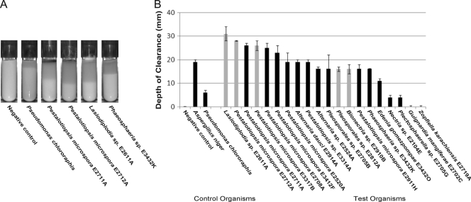
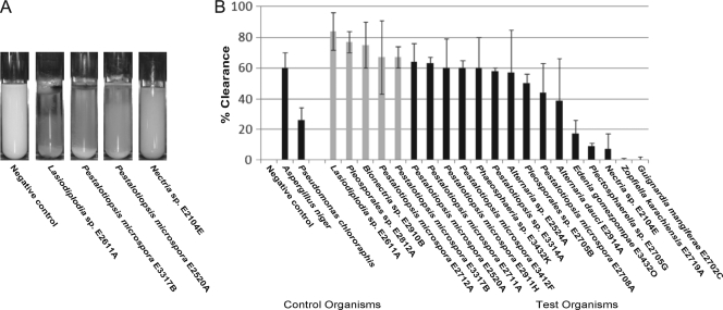
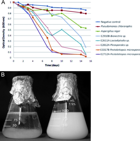
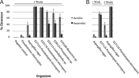
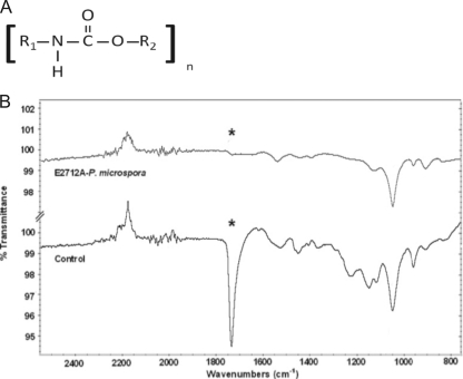
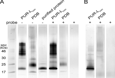
Bioremediation is an important approach to waste reduction that relies on biological processes to break down a variety of pollutants. This is made possible by the vast metabolic diversity of the microbial world. To explore this diversity for the breakdown of plastic, we screened several dozen endophytic fungi for their ability to degrade the synthetic polymer polyester polyurethane (PUR). Several organisms demonstrated the ability to efficiently degrade PUR in both solid and liquid suspensions. Particularly robust activity was observed among several isolates in the genus Pestalotiopsis, although it was not a universal feature of this genus. Two Pestalotiopsis microspora isolates were uniquely able to grow on PUR as the sole carbon source under both aerobic and anaerobic conditions. Molecular characterization of this activity suggests that a serine hydrolase is responsible for degradation of PUR. The broad distribution of activity observed and the unprecedented case of anaerobic growth using PUR as the sole carbon source suggest that endophytes are a promising source of biodiversity from which to screen for metabolic properties useful for bioremediation.


Figures

Fig. 1.
Fig. 1.
Example of PUR-A plates initially used to screen for polyurethane-degrading activity after 2 weeks of fungal growth. (A) Negative control. (B) Pleosporales sp. strain E2705B.
Fig. 2.
Fig. 2.
(A) Examples of PUR-A solid agar cultures at 2 weeks after inoculation, including Pseudomonas chlororaphis as a positive control. (B) Fungal cultures were grown in triplicate on solid PUR-A medium for 2 weeks. Depth-of-clearance measurements were made after 2 weeks of growth. The positive-control test organisms, Pseudomonas chlororaphis and Aspergillus niger, are represented at the left. The five highlighted organisms (Lasiodiplodia sp. strain E2611A, Pestalotiopsis microspora E2712A, Pestalotiopsis microspora E3317B, Pleosporales sp. strain E2812A, and Bionectria sp. strain E2910B) were selected for further screening. Error bars represent the standard deviation for each data set.
Fig. 3.
Fig. 3.
(A) Examples of PUR-L liquid cultures at 2 weeks after inoculation. (B) Fungal cultures were grown in triplicate in liquid PUR-L medium containing Impranil DLF for 2 weeks. Percent clearance was determined by a decrease in light scattering at 600 nm as measured by UV-visible spectrophotometry. Percent clearance measurements were taken after 2 weeks of growth. All data were normalized to the negative control. The positive-control test organisms, Pseudomonas chlororaphis and Aspergillus niger, are represented at the left. The 5 highlighted organisms (Lasiodiplodia sp. strain E2611A, Pestalotiopsis microspora E2712A, Pestalotiopsis microspora E3317B, Pleosporales sp. strain E2812A, and Bionectria sp. strain E2910B) were selected for further screening. Error bars represent the standard deviation for each data set.
Fig. 4.
Fig. 4.
(A) Degradation of PUR as a sole carbon source by the five most active organisms was monitored over a 16-day time course. Cultures containing PUR-Lmin medium with Impranil DLN as the sole carbon source were inoculated with washed fungal inoculums. The assay was performed in duplicate, and the values shown represent the averages for the two cultures at each time point. Aspergillus niger (green) and Pseudomonas chlororaphis (red) served as the positive controls. (B) Pestalotiopsis microspora E2712A (left) degrading PUR Impranil DLN as a sole carbon source after a 16-day time course. The remaining material in the flask is the result of fungal growth.
Fig. 5.
Fig. 5.
(A) PUR degradation measurements of fungal cultures grown under aerobic and anaerobic conditions. Triplicate sets of fungal cultures were grown in PUR-Lmin medium containing Impranil DLN as the sole carbon medium under aerobic or anaerobic conditions. At the end of 2 weeks of growth, samples were removed from the sealed chamber and the percent clearance of the PUR in solution for each set of cultures was measured by decreased light scattering at a wavelength of 600 nm using UV-visible spectrophotometry The results for the controls Pseudomonas chlororaphis and Aspergillus niger are shown at the left. Two strains of Pestalotiopsis microspora (E2712A and E3317B) maintained polyurethanase activity when grown under anaerobic conditions, while Pseudomonas chlororaphis, Aspergillus niger, Lasiodiplodia sp. strain E2611A, Pleosporales sp. strain E2812A, and Bionectria sp. strain E2910B all showed significant reductions in the magnitude of PUR degradation activity. Error bars represent the standard deviation for each data set. (B) The rates of degradation were compared by measurement in triplicate of cultures of Aspergillus niger and Pestalotiopsis microspora E2712A after 1 week and 2 weeks of growth. Pestalotiopsis microspora E2712A shows equivalent rates of degradation activity under aerobic and anaerobic conditions. Error bars represent the standard deviation for each data set.
Fig. 6.
Fig. 6.
(A) General chemical structure of the polyurethane molecule. R1 and R2 connect subsequent urethane monomers within the polymeric molecule. (B) Infrared spectra of PUR liquid medium containing Impranil DLN taken after 6 days of incubation with the fungus Pestalotiopsis microspora. The sample spectrum (top) and control spectrum (bottom) are shown together with a common scale. The strong peak denoted by an asterisk at 1,735 cm−1 in the control corresponds to the ester C=O stretch in the ethyl carbamate of the urethane motif. This peak disappears after degradation of the ester linkage by the polyurethanase enzyme.
Fig. 7.
Fig. 7.
Probing for serine hydrolases in crude protein from cell-free filtrate. SDS-PAGE analysis of proteins from a crude cell-free filtrate of Pestalotiopsis microspora E2712A grown in PUR-Lmin medium and PDB rich medium is shown. Cell-free filtrates were incubated with serine hydrolase activity-based probes prior to loading. (A) Protein bands were visualized by silver staining. (B) Labeled protein bands were visualized using a fluorescence scanner. This analysis shows that an approximately 21-kDa protein expressed exclusively under the induced PUR-Lmin growth condition is efficiently labeled by the serine hydrolase activity-based probe.

References

    1. Akutsu Y., Nakajima-Kambe T., Nomura N., Nakahara T. 1998. Purification and properties of a polyester-polyurethane degrading enzyme from Comamonas acidovorans TB-35. Appl. Environ. Microbiol. 64:62–67 - PMC - PubMed
    1. Allen B. A., Hilliard N. P., Howard G. T. 1999. Purification and characterization of a soluble polyurethane degrading enzyme from Comamonas acidovorans. Int. Biodeterior. Biodegrad. 43:37–41
    1. Bacon C., White J. 2000. Microbial endophytes. Marcel Dekker, New York, NY
    1. Cosgrove L., McGeechan P. L., Robson G. D., Handley P. S. 2007. Fungal communities associated with degradation of polyester polyurethane in soil. Appl. Environ. Microbiol. 73:5817–5824 - PMC - PubMed
    1. Crabbe J. R., Campbell J. R., Thompson L., Walz S. L., Schultz W. W. 1994. Biodegradation of a colloidal ester-based polyurethane by soil fungi. Int. Biodeterior. Biodegrad. 33:103–113

Publication types

MeSH terms