. 2011 Sep;77(17):5945-54.
doi: 10.1128/AEM.03022-10. Epub 2011 Jul 15.

Novel virulent and broad-host-range Erwinia amylovora bacteriophages reveal a high degree of mosaicism and a relationship to Enterobacteriaceae phages

Affiliations

Novel virulent and broad-host-range Erwinia amylovora bacteriophages reveal a high degree of mosaicism and a relationship to Enterobacteriaceae phages

Yannick Born et al. Appl Environ Microbiol. 2011 Sep.

Abstract

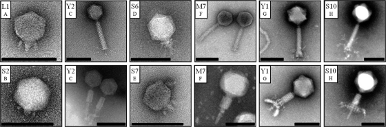
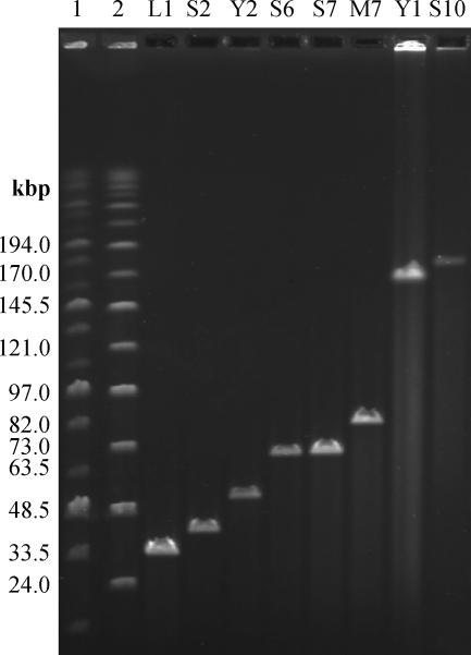
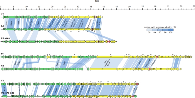
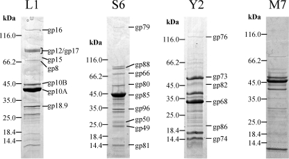
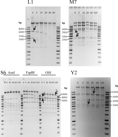
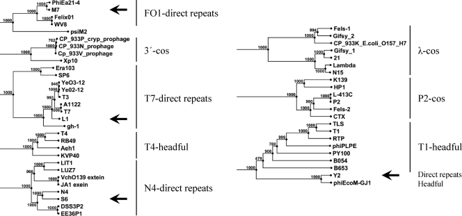
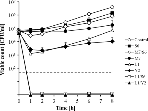
A diverse set of 24 novel phages infecting the fire blight pathogen Erwinia amylovora was isolated from fruit production environments in Switzerland. Based on initial screening, four phages (L1, M7, S6, and Y2) with broad host ranges were selected for detailed characterization and genome sequencing. Phage L1 is a member of the Podoviridae, with a 39.3-kbp genome featuring invariable genome ends with direct terminal repeats. Phage S6, another podovirus, was also found to possess direct terminal repeats but has a larger genome (74.7 kbp), and the virus particle exhibits a complex tail fiber structure. Phages M7 and Y2 both belong to the Myoviridae family and feature long, contractile tails and genomes of 84.7 kbp (M7) and 56.6 kbp (Y2), respectively, with direct terminal repeats. The architecture of all four phage genomes is typical for tailed phages, i.e., organized into function-specific gene clusters. All four phages completely lack genes or functions associated with lysogeny control, which correlates well with their broad host ranges and indicates strictly lytic (virulent) lifestyles without the possibility for host lysogenization. Comparative genomics revealed that M7 is similar to E. amylovora virus ΦEa21-4, whereas L1, S6, and Y2 are unrelated to any other E. amylovora phage. Instead, they feature similarities to enterobacterial viruses T7, N4, and ΦEcoM-GJ1. In a series of laboratory experiments, we provide proof of concept that specific two-phage cocktails offer the potential for biocontrol of the pathogen.


Figures

Fig. 1.
Fig. 1.
Electron micrographs of negatively stained Erwinia amylovora phages. Phages were classified into the Podoviridae (L1, S2, S6, and S7) and Myoviridae (Y2, M7, Y1, and S10) families. From the latter, virions with both contracted and uncontracted tails are shown. Letters A to H indicate phage subtypes. Scale bars represent 100 nm.
Fig. 2.
Fig. 2.
Pulsed-field gel electrophoresis analysis of full-length Erwinia amylovora phage genomes (as indicated). Lanes 1 and 2 include MidRange PFGE markers I and II (NEB), respectively. Sizes are as indicated.
Fig. 3.
Fig. 3.
Genome maps of phages L1, S6, and Y2 and alignment with related phages. Genes were classified into functional clusters based on database comparisons, peptide fingerprinting, and their locations on the genome. They are represented in green (early genes), yellow (late genes), and red (lysis genes). Shading indicates the degree of amino acid sequence identity of gene products with an identity of >20%. Putative functions of selected genes are indicated. Gene products that were identified using mass spectrometry are marked with an asterisk.
Fig. 4.
Fig. 4.
Analysis of virion proteins of phages L1, S6, Y2, and M7 by SDS-PAGE. Molecular mass markers are indicated on the left of each gel segment. Identification of individual protein bands was performed by peptide fingerprinting and mass spectrometry. The corresponding gene product names are indicated on the right (except for phage M7).
Fig. 5.
Fig. 5.
Analysis of phage genome physical structures. Shown are fragment patterns of phage DNA, following time-limited BAL31 treatment and subsequent digestion with different endonucleases. Phage L1 was digested with MfeI, M7 with FspBI, S6 with three different restriction endonucleases, and Y2 with OliI. Arrows indicate terminal fragments, which are progressively shortened by the BAL31 nuclease. The numbered arrow in the lower left panel (S6) indicates the second-to-last fragment, which is shortened only after the terminal fragment is completely degraded. Incubation times with BAL31 in minutes are indicated at the top of each gel.
Fig. 6.
Fig. 6.
Phylogenetic analysis of large terminase subunits of phages L1, M7, S6, and Y2 (indicated by arrows) and comparison to other phages with known packaging mechanisms (11, 30) as indicated. The tree was generated from an alignment (gap open cost, 10; gap extension cost, 1; end gap cost, free) using the neighbor-joining method with 1,000 bootstrap replicates (CLC Main Workbench 5).
Fig. 7.
Fig. 7.
Viable counts of E. amylovora CFBP1430 after infection with single phages or two-phage cocktails. The values indicate the means of results from three independent trials, including standard deviations. The dashed line indicates the detection limit of 50 CFU/ml.

References

    1. Ackermann H.-W. 1996. Frequency of morphological phage descriptions in 1995. Arch. Virol. 141:209–218 - PubMed
    1. Adams M. H. 1959. Bacteriophages. Interscience Publishers Inc., New York, NY
    1. Altschul S. F., Gish W., Miller W., Myers E. W., Lipman D. J. 1990. Basic local alignment search tool. J. Mol. Biol. 215:403–410 - PubMed
    1. Balogh B., Canteros B. I., Stall R. E., Jones J. B. 2008. Control of citrus canker and citrus bacterial spot with bacteriophages. Plant Dis. 92:1048–1052 - PubMed
    1. Balogh B., Jones J. B., Iriarte F. B., Momol M. T. 2010. Phage therapy for plant disease control. Curr. Pharm. Biotechnol. 11:48–57 - PubMed

Publication types

MeSH terms

Associated data

LinkOut - more resources