

Blast-induced phenotypic switching in cerebral vasospasm
- PMID: 21765001
- PMCID: PMC3150891
- DOI: 10.1073/pnas.1105860108
Blast-induced phenotypic switching in cerebral vasospasm
Abstract
Vasospasm of the cerebrovasculature is a common manifestation of blast-induced traumatic brain injury (bTBI) reported among combat casualties in the conflicts in Afghanistan and Iraq. Cerebral vasospasm occurs more frequently, and with earlier onset, in bTBI patients than in patients with other TBI injury modes, such as blunt force trauma. Though vasospasm is usually associated with the presence of subarachnoid hemorrhage (SAH), SAH is not required for vasospasm in bTBI, which suggests that the unique mechanics of blast injury could potentiate vasospasm onset, accounting for the increased incidence. Here, using theoretical and in vitro models, we show that a single rapid mechanical insult can induce vascular hypercontractility and remodeling, indicative of vasospasm initiation. We employed high-velocity stretching of engineered arterial lamellae to simulate the mechanical forces of a blast pulse on the vasculature. An hour after a simulated blast, injured tissues displayed altered intracellular calcium dynamics leading to hypersensitivity to contractile stimulus with endothelin-1. One day after simulated blast, tissues exhibited blast force dependent prolonged hypercontraction and vascular smooth muscle phenotype switching, indicative of remodeling. These results suggest that an acute, blast-like injury is sufficient to induce a hypercontraction-induced genetic switch that potentiates vascular remodeling, and cerebral vasospasm, in bTBI patients.
Conflict of interest statement
The authors declare no conflict of interest.
Figures
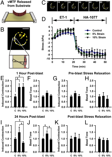
 ). (C-D) vMTFs were serially stimulated with ET-1 and HA-1077. (C) Single vMTF during experimental protocol. (
 ). (D) Temporal change in vMTF stress due to serial stimulation, 1 h after simulated blast. (E–F) Normalized ET-1 induced contraction and basal tone 1 h after simulated blast. (G–H) Normalized induced contraction and basal tone 1 h after simulated blast for tissues pretreated with ROCK inhibitor. (I–J) Normalized induced contraction and basal tone 24 h after simulated blast. (K–L) Normalized induced contraction and basal tone 24 h after simulated blast for tissues treated ROCK inhibitor immediately following the blast. All graphs: mean + /-SEM.
References
-
- Warden DL, French L. Traumatic brain injury in the war zone. N Engl J Med. 2005;353:633–634. - PubMed
-
- Bhattacharjee Y. Neuroscience. shell shock revisited: solving the puzzle of blast trauma. Science. 2008;319:406–408. - PubMed
-
- Ling G, Bandak F, Armonda R, Grant G, Ecklund J. Explosive blast neurotrauma. J Neurotrauma. 2009;26:815–825. - PubMed
-
- Borel CO, et al. Possible role for vascular cell proliferation in cerebral vasospasm after subarachnoid hemorrhage. Stroke. 2003;34:427–432. - PubMed
-
- Zhang ZD, Macdonald RL. Contribution of the remodeling response to cerebral vasospasm. Neurol Res. 2006;28:713–720. - PubMed
Publication types
MeSH terms
Substances
LinkOut - more resources
Full Text Sources
Research Materials
