. 2011 Dec;39(12):2711-21.
doi: 10.1097/CCM.0b013e3182284a5f.

Induction of cellular antioxidant defense by amifostine improves ventilator-induced lung injury

Affiliations

Induction of cellular antioxidant defense by amifostine improves ventilator-induced lung injury

Panfeng Fu et al. Crit Care Med. 2011 Dec.

Abstract

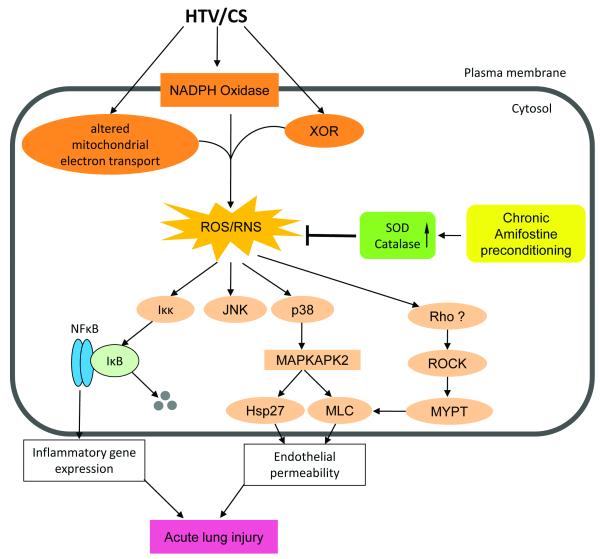
Objectives: To test the hypothesis that preconditioning animals with amifostine improves ventilator-induced lung injury via induction of antioxidant defense enzymes. Mechanical ventilation at high tidal volume induces reactive oxygen species production and oxidative stress in the lung, which plays a major role in the pathogenesis of ventilator-induced lung injury. Amifostine attenuates oxidative stress and improves lipopolysaccharide-induced lung injury by acting as a direct scavenger of reactive oxygen and nitrogen species. This study tested effects of chronic amifostine administration on parameters of oxidative stress, lung barrier function, and inflammation associated with ventilator-induced lung injury.

Design: Randomized and controlled laboratory investigation in mice and cell culture.

Setting: University laboratory.

Subjects: C57BL/6J mice.

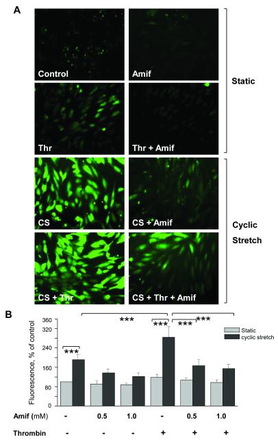
Interventions: Mice received once-daily dosing with amifostine (10-100 mg/kg, intraperitoneal injection) 3 days consecutively before high tidal volume ventilation (30 mL/kg, 4 hrs) at day 4. Pulmonary endothelial cell cultures were exposed to pathologic cyclic stretching (18% equibiaxial stretch) and thrombin in a previously verified two-hit model of in vitro ventilator-induced lung injury.

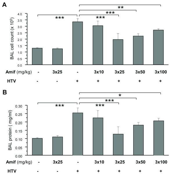
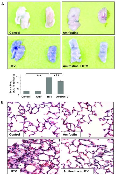
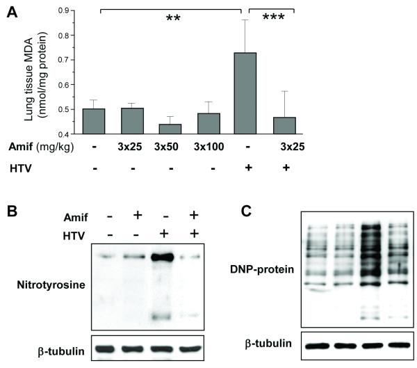
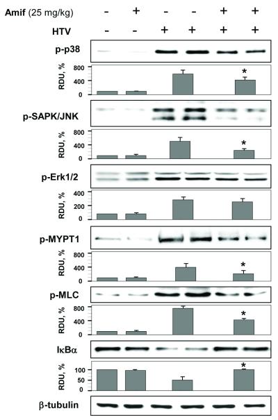
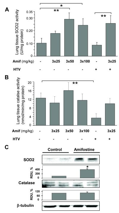
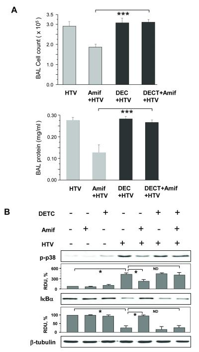
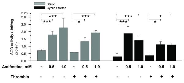
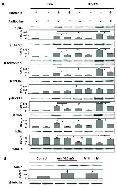
Measurements and main results: Three-day amifostine preconditioning before high tidal volume attenuated high tidal volume-induced protein and cell accumulation in the alveolar space judged by bronchoalveolar lavage fluid analysis, decreased Evans Blue dye extravasation into the lung parenchyma, decreased biochemical parameters of high tidal volume-induced tissue oxidative stress, and inhibited high tidal volume-induced activation of redox-sensitive stress kinases and nuclear factor-kappa B inflammatory cascade. These protective effects of amifostine were associated with increased superoxide dismutase 2 expression and increased superoxide dismutase and catalase enzymatic activities in the animal and endothelial cell culture models of ventilator-induced lung injury.

Conclusions: Amifostine preconditioning activates lung tissue antioxidant cell defense mechanisms and may be a promising strategy for alleviation of ventilator-induced lung injury in critically ill patients subjected to extended mechanical ventilation.


Figures

Figure 1
Figure 1. Chronic amifostine preconditioning attenuates high tidal volume ventilation-induced lung injury
A and B - Mice were pretreated with amifostine for 3 days followed by ventilation at HTV. Control animals were treated with vehicle (PBS) or amifostine alone. Cell count (A) and protein concentration (B) were measured in bronchoalveolar lavage fluid taken from control and experimental animals. Data are presented as mean ± SD; n=4 in vehicle and amifostine groups; n=5 in groups of amifostine + HTV; n=10 in HTV group; *p<0.05, **p<0.01, ***p<0.001.
Figure 2
Figure 2. Chronic amifostine preconditioning alleviates lung vascular permeability and cellular infiltration induced by high tidal volume mechanical ventilation
Mice were pretreated with vehicle or amifostine (25 mg/kg) for 3 days followed by ventilation at HTV. Control animals were treated with vehicle (PBS) or amifostine alone. A - Lung vascular permeability was assessed by Evans blue accumulation in the lungs. The panels show right and left lungs respectively, which were excised after perfusion to remove blood and imaged. Bar graph depicts quantitative spectrophotomertic analysis of Evans blue-labeled albumin content in the lung tissues. Data are presented as mean ± SD; n=4-6 mice per group; ***p<0.001. B - Lung specimen from control and experimental animals were stained with hematoxylin and eosin. Images are representative of 4-6 lung specimen for each condition; ×40 magnification.
Figure 3
Figure 3. Amifostine attenuates lung oxidative stress by HTV
A - Measurements of malone dialdehyde (MDA) content in control and HTV-exposed lungs with or without amifostine preconditioning. Data are presented as mean ± SD; n=4 in vehicle group; n=6 in the other groups; **p<0.01, ***p<0.001. B - levels of nitrotyrosinated proteins, and C - DNP-derivatized oxidized proteins in lung tissue samples as indices of oxidative stress were analyzed by western blot in control and amifostine-peconditioned (25 mg/kg, three daily i.p. injections) lungs after HTV exposure. Shown are representative results of 5 experiments.
Figure 4
Figure 4. Amifostine inhibits HTV-induced MAP kinase signaling and IκBα degradation in mouse lungs
Lungs from control or amifostine-preconditioned (25 mg/kg, three daily i.p. injections) mice were subjected to HTV. Control animals were treated with vehicle (PBS) or amifostine alone. Phosphorylation of p38, JNK, Erk-1,2, MYPT and MLC, and degradation of IκBα in tissue samples was analyzed by western blot. Tissue samples from HTV-exposed animals with and without amifostine pretreatment are presented in duplicates. Equal protein loading was confirmed by probing of membranes with β-tubulin antibodies. Results of densitometry normalized to β-tubulin signal are shown as mean±SD; n=6 per condition. Bars depict marker phospho-protein levels in tissue samples corresponding to control group without amifostine pretreatment; HTV; and HTV+amifostine groups. *p<0.05 as compared to HTV.
Figure 5
Figure 5. Effect of amifostine on the enzyme activities of SOD and catalse and their expression in lung tissue
Mice were pretreated with vehicle (PBS) or amifostine for 3 days followed by ventilation at HTV. A and B - Measurements of SOD (A) or catalase (B) activity. Data are presented as mean ± SD; n=4-6 mice per each dose group; *p<0.05, **p<0.01. C - Protein expression of SOD2 and catalase in control and amifostine-preconditioned lungs was analyzed by western blot of tissue samples. The expression levels of each protein was quantified by densitometry and normalized to β-tubulin signal. Tissue samples are presented in duplicates. Results shown as mean±SD; n=6 for each group; *p<0.05.
Figure 6
Figure 6. SOD inhibitor DECT relieves protective effect of amifostine against HTV-induced lung injury and blocking of HTV-induced stress signaling
Mice were preconditioned with amifostine (25 mg/kg, three daily injections) with or without DETC injection prior to HTV. Control animals were treated with vehicle (PBS). A - Measurements of cell counts and protein content in BAL samples. Values are mean ± SD, n=4 for each group; ***p<0.001. B - Phosphorylation of p38 MAP kinase and degradation of IκBα was analyzed by western blot and quantified by densitometry. Results of densitometry shown as mean±SD; n=4 for each group; *p<0.05.
Figure 7
Figure 7. Amifostine inhibits cyclic stretch- and thrombin-induced ROS production in human pulmonary EC
Control and amifostine-preconditioned (Amif) HPAEC grown on BioFlex plates were exposed to 18% cyclic stretch (CS) followed by thrombin (Thr) stimulation or left under static conditions. To detect ROS, 30 min prior to termination of the experiment cells were incubated with fluorescent dye for ROS, dichlorodihydrofluorescein (DCFDA, 10 μM) under continuing CS stimulation. A -Cells were imaged using inverted fluorescent microscope. B - Fluorescence intensity determined in vehicle control cells was taken as 100%. Results of 6 images from the entire cell monolayer were averaged and have been reproduced in 3 independent experiments. Results are presented as mean ± SD; ***p<0.001.
Figure 8
Figure 8. Amifostine pretreatment increases SOD activity in human pulmonary endothelial cells
HPAEC were pretreated with vehicle (PBS) or amifostine for 3 days followed by 18% CS with or without thrombin challenge. Control cells were left under static conditions. Measurements of SOD activity were performed in control and stimulated cells. Data are presented as mean ± SD; n=3 for each group; *p<0.05; ***p<0.001.
Figure 9
Figure 9. Amifostine pretreatment attenuates cyclic stretch- and thrombin-induced activation of stress cascades and barrier disruptive signaling
HPAEC were preconditioned with vehicle (PBS) or amifostine followed by 18% CS with or without thrombin treatment, or left static. A - Effect of amifostine preconditioning on activation of oxidative stress sensitive pathways was analyzed by western blot with specific antibodies and quantitative densitometry analysis. Results of densitometry shown as mean±SD; n=4 for each group; *p<0.05. B - SOD2 expression levels in HPAEC were quantified by densitometry and normalized to β-tubulin. Results shown as mean±SD; n=6 for each group; *p<0.05.
Figure 10
Figure 10. Attenuation of ventilator induced lung injury by amifostine
Mechanical stretch associated with high tidal volume mechanical ventilation induces reactive oxygen species (ROS) and reactive nitrogen species (RNS) production, leading to activation of redox-sensitive stress kinases (JNK and p38 MAPK) and NFkB signaling cascade leading to expression of pro-inflammatory cytokines, cytoskeletal remodeling and disruption of endothelial monolayer integrity. Amifostine attenuates oxidative stress induced by HTV through upregulation of SOD protein expression and stimulation of SOD and catalase enzymatic activities. XOR – xanthine oxidoreductase; SOD – superoxide dismutase; MAPKAPK2 – MAPK-activated protein kinase 2 ; Hsp27 – heat shock protein 27; MLC – myosin light chains; ROCK – Rho associated kinase; MYPT – myosin protein phosphatase.

Comment in

Similar articles

Cited by

References

    1. Rubenfeld GD, Caldwell E, Peabody E, Weaver J, Martin DP, Neff M, et al. Incidence and outcomes of acute lung injury. N Engl J Med. 2005 Oct 20;353(16):1685–93. - PubMed
    1. Ware LB, Matthay MA. The acute respiratory distress syndrome. N Engl J Med. 2000 May 4;342(18):1334–49. - PubMed
    1. Ranieri VM, Suter PM, Tortorella C, De Tullio R, Dayer JM, Brienza A, et al. Effect of mechanical ventilation on inflammatory mediators in patients with acute respiratory distress syndrome: a randomized controlled trial. Jama. 1999 Jul 7;282(1):54–61. - PubMed
    1. Tremblay L, Valenza F, Ribeiro SP, Li J, Slutsky AS. Injurious ventilatory strategies increase cytokines and c-fos m-RNA expression in an isolated rat lung model. J Clin Invest. 1997 Mar 1;99(5):944–52. - PMC - PubMed
    1. Villar J, Flores C, Mendez-Alvarez S. Genetic susceptibility to acute lung injury. Crit Care Med. 2003 Apr;31(4 Suppl):S272–5. - PubMed

Publication types