. 2011 Jul 17;476(7358):92-5.
doi: 10.1038/nature10206.

Photoentrainment and pupillary light reflex are mediated by distinct populations of ipRGCs

Affiliations

Photoentrainment and pupillary light reflex are mediated by distinct populations of ipRGCs

S-K Chen et al. Nature. .

Abstract

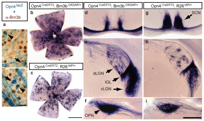
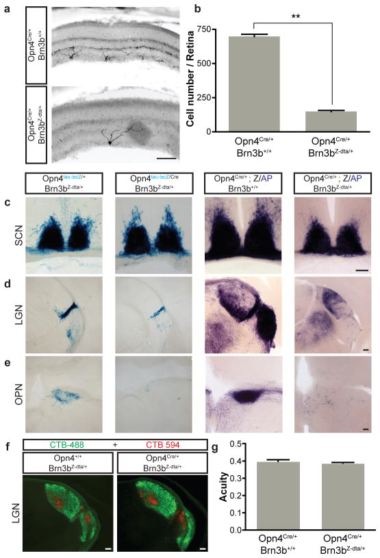
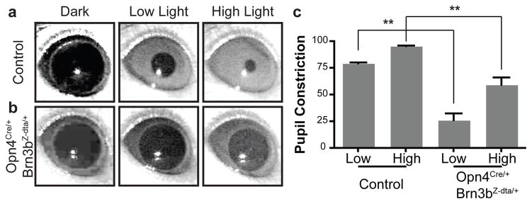
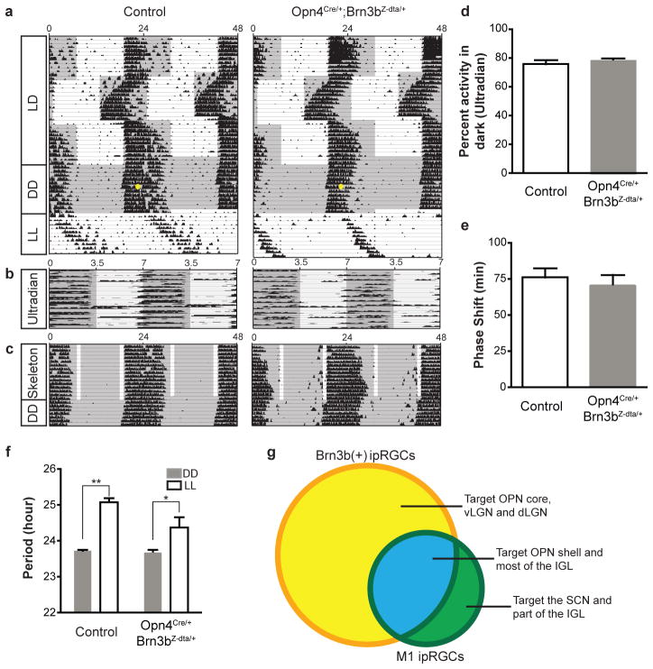
Intrinsically photosensitive retinal ganglion cells (ipRGCs) express the photopigment melanopsin and regulate a wide array of light-dependent physiological processes. Genetic ablation of ipRGCs eliminates circadian photoentrainment and severely disrupts the pupillary light reflex (PLR). Here we show that ipRGCs consist of distinct subpopulations that differentially express the Brn3b transcription factor, and can be functionally distinguished. Brn3b-negative M1 ipRGCs innervate the suprachiasmatic nucleus (SCN) of the hypothalamus, whereas Brn3b-positive ipRGCs innervate all other known brain targets, including the olivary pretectal nucleus. Consistent with these innervation patterns, selective ablation of Brn3b-positive ipRGCs severely disrupts the PLR, but does not impair circadian photoentrainment. Thus, we find that molecularly distinct subpopulations of M1 ipRGCs, which are morphologically and electrophysiologically similar, innervate different brain regions to execute specific light-induced functions.


Conflict of interest statement

The authors declare no competing financial interests.

Figures

Figure 1
Figure 1. Co-expression of Melanopsin and Brn3b defines a specific set of ipRGCs
a, Retinal flat mounts from Opn4tau-LacZ/+ mice stained with anti-Brn3b antibody (brown) and X-gal staining (blue) show Brn3b-positive (arrowheads; 140 Brn3b-positive ipRGCs from 988 lacZ+ cells, n=5) and Brn3b-negative (arrows), M1 ipRGCs. b–i, AP histochemistry of retina (b and c) and coronal brain sections (d–i) from Opn4CreERT2/+;Brn3bCKOAP/+ mice (b, d–f), or from Opn4CreERT2/+;R26IAP/+ mice (c, g–i). Suprachiasmatic region shows partial innervation in Opn4CreERT2/+;Brn3bCKOAP/+ mice (d), compared to full innervation of the SCN in Opn4CreERT2/+;R26IAP/+ mice (g). Both mouse lines show significant ipRGC projections to the IGL and vLGN, and sparse innervation to the dLGN (e and h) and intense labeling of the OPN (f and i). Scale bars are 25 μm (a), 1 mm (b and c), and 400 μm (d–i).
Figure 2
Figure 2. Genetic ablation of Brn3b-positive ipRGCs does not impair targeting to the SCN
a, Melanopsin immunofluorescence reveals a reduction in ipRGC numbers in pn4Cre/+;Brn3bZ-dta/+ retina compared to control (Opn4Cre/+;Brn3b+/+). b, Quantification of surviving melanopsin-positive cells in Opn4Cre/+;Brn3bZ-dta/+ (149.8±8.65 cells/retina; n=4) and control (698.8±16.85 cells/retina; n=4) mice. c–e, Coronal brain sections of pn4tau-LacZ/+;Brn3bZ-dta/+ and Opn4Cre/tau-LacZ;Brn3bZ-dta/+ (c–e, left two panels), and pn4Cre/+;Brn3b+/+;Z/AP and Opn4Cre/+;Brn3bZ-dta/+;Z/AP (c–e, right two panels) mice using X-gal (c–e, left two panels) or AP histochemistry (c–e, right two panels). Sections show SCN (c), LGN (d), and OPN (e). i, Labeling of all RGCs with Alexa Fluor 594- and 488-conjugated Cholera toxin B in the left eye (red) and the right eye (green), respectively shows normal brain targeting to image forming regions. j, Visual acuity was the same between Opn4+/+;Brn3bZ-dta/+ (n=5) and Opn4Cre/+;Brn3bZ-dta/+ mice (n=6). Scale bars are 100 μm. Error bars represent SEMs.
Figure 3
Figure 3. Opn4Cre/+;Brn3Z-dta/+ mice show severe deficits in the pupillary light reflex PLR)
a–b, Representative images of PLR from control and Opn4Cre/+;Brn3bZ-dta/+ mice. Left panels show pupils under dark conditions, middle panels show pupils under low light intensity (22 μW/cm2) and right panels show pupils under high light intensity (5.66 mW/cm2). c, Quantification of PLR data from control (n=5) and Opn4Cre/+;Brn3bZ-dta/+ n=6) animals. ** indicates p<0.01 with 1-way ANOVA. Error bars represent SEMs.
Figure 4
Figure 4. Opn4Cre/+;Brn3bZ-dta/+ mice show normal circadian photoentrainment
a–c, Representative actograms from control and Opn4Cre/+;Brn3bZ-dta/+ animals under a series of lighting paradigms: a, 12:12 hours LD cycle, “jet-lag”, constant darkness (DD), and constant light (LL); b, Ultradian 3.5:3.5 hours cycles; c, skeleton photoperiod. The gray background indicates darkness and the yellow dot indicates the 15 minutes light pulse at CT 16. Opn4Cre/+;Brn3bZ-dta/+ animals have similar photoentrainment to controls with minor deficits in period lengthening. d, Percent activity in the dark portion of the ultradian cycle shows no significant difference between the genotypes. e, Quantification of phase shifts shows no significant differences between the two groups. f, Quantification of circadian period from the two groups under constant dark and constant light conditions. Both groups of animals show significant period lengthening under constant light. g) Venn diagram showing Brn3b-positive ipRGCs in yellow and Brn3b negative ipRGCs in green (full description is provided in supplementary table 1). ** indicates p<0.01, * indicates p<0.05 using student’s t-test. Error bars represent SEMs.

References

    1. Berson DM, Dunn FA, Takao M. Phototransduction by retinal ganglion cells that set the circadian clock. Science. 2002;295 (5557):1070–1073. - PubMed
    1. Ecker JL, et al. Melanopsin-expressing retinal ganglion-cell photoreceptors: cellular diversity and role in pattern vision. Neuron. 2010;67 (1):49–60. - PMC - PubMed
    1. Gooley JJ, Lu J, Fischer D, Saper CB. A broad role for melanopsin in nonvisual photoreception. J Neurosci. 2003;23 (18):7093–7106. - PMC - PubMed
    1. Hannibal J, Fahrenkrug J. Melanopsin containing retinal ganglion cells are light responsive from birth. Neuroreport. 2004;15 (15):2317–2320. - PubMed
    1. Hattar S, Liao HW, Takao M, Berson DM, Yau KW. Melanopsin-containing retinal ganglion cells: architecture, projections, and intrinsic photosensitivity. Science. 2002;295 (5557):1065–1070. - PMC - PubMed

Publication types

MeSH terms