. 2012 Feb 16;31(7):884-96.
doi: 10.1038/onc.2011.288. Epub 2011 Jul 18.

The interaction between caveolin-1 and Rho-GTPases promotes metastasis by controlling the expression of alpha5-integrin and the activation of Src, Ras and Erk

Affiliations
Free PMC article

The interaction between caveolin-1 and Rho-GTPases promotes metastasis by controlling the expression of alpha5-integrin and the activation of Src, Ras and Erk

E Arpaia et al. Oncogene. .
Free PMC article

Abstract

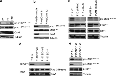
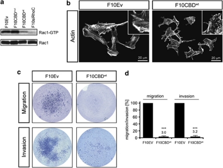
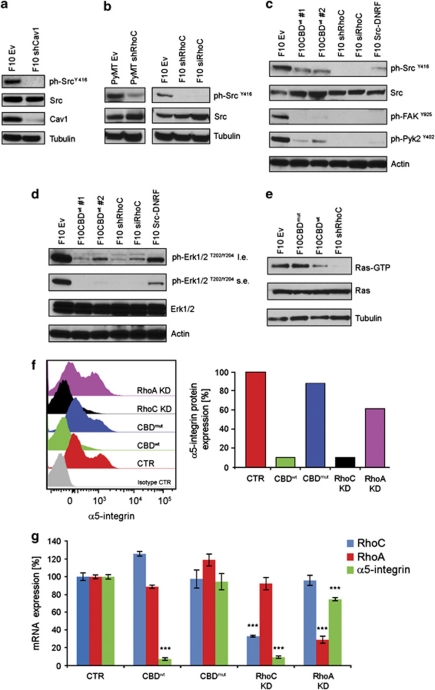
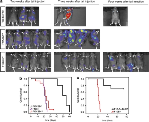
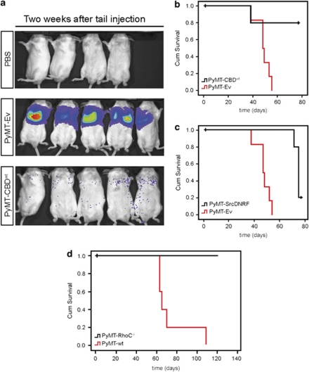
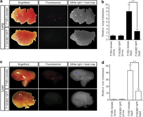
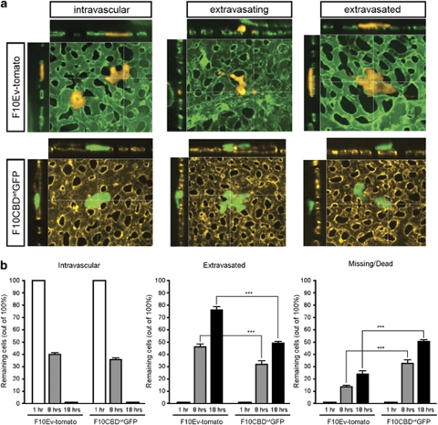
Proteins containing a caveolin-binding domain (CBD), such as the Rho-GTPases, can interact with caveolin-1 (Cav1) through its caveolin scaffold domain. Rho-GTPases are important regulators of p130(Cas), which is crucial for both normal cell migration and Src kinase-mediated metastasis of cancer cells. However, although Rho-GTPases (particularly RhoC) and Cav1 have been linked to cancer progression and metastasis, the underlying molecular mechanisms are largely unknown. To investigate the function of Cav1-Rho-GTPase interaction in metastasis, we disrupted Cav1-Rho-GTPase binding in melanoma and mammary epithelial tumor cells by overexpressing CBD, and examined the loss-of-function of RhoC in metastatic cancer cells. Cancer cells overexpressing CBD or lacking RhoC had reduced p130(Cas) phosphorylation and Rac1 activation, resulting in an inhibition of migration and invasion in vitro. The activity of Src and the activation of its downstream targets FAK, Pyk2, Ras and extracellular signal-regulated kinase (Erk)1/2 were also impaired. A reduction in α5-integrin expression, which is required for binding to fibronectin and thus cell migration and survival, was observed in CBD-expressing cells and cells lacking RhoC. As a result of these defects, CBD-expressing melanoma cells had a reduced ability to metastasize in recipient mice, and impaired extravasation and survival in secondary sites in chicken embryos. Our data indicate that interaction between Cav1 and Rho-GTPases (most likely RhoC but not RhoA) promotes metastasis by stimulating α5-integrin expression and regulating the Src-dependent activation of p130(Cas)/Rac1, FAK/Pyk2 and Ras/Erk1/2 signaling cascades.


Figures

Figure 1
Figure 1
RhoC and Cav1 regulate p130Cas phosphorylation. (a) Correlation between Cav1 and ph-p130Cas. The indicated clones of B16 melanoma cells were evaluated by immunoblotting (IB) for levels of Cav1 and p130Cas phosphorylation at Y410 or Y165. (b) Transient silencing (two independent clones #1, #2) of Cav1 expression reduces p130CasY410 phosphorylation. (c) RhoC controls ph-p130Cas levels. F10 cells stably expressing siRhoC or shRhoC were analyzed by IB to detect ph-p130Cas and Cav1. The ph-p130Cas levels are also reduced in PyMT cells expressing shRhoC. (d) CBDwt inhibits the binding of Rho-GTPases to Cav1. Active Rho-GTPases were pulled-down with agarose beads GST-Rhoteckin-RBD-conjugated (RBD: Rho Binding Domain) and Cav1 was detected by IB. (e) CBDwt expression reduces ph-p130Cas. The levels of p130Cas and p130Cas phosphorylated at Y410 or Y165 were assessed by IB in F10Ev (control F10 cells expressing empty vector) and two independent clones of F10 cells overexpressing CBDwt (F10CBDwt #1, #2).
Figure 2
Figure 2
Effects of disrupting the Cav1–Rho-GTPase interaction. (a) Decreased Rac1 activation in the presence of CBDwt or absence of RhoC. Active Rac1 (Rac1-GTP) was pulled down with GST-PAK-PDB-conjugated agarose beads (PAK: p21-activated kinase 1; PDB: p21-binding domain) from extracts of F10 cells expressing Ev, CBDmut, CBDwt, siRhoC. Rac1 was detected by IB. (b) CDBwt expression alters cytoskeletal remodeling. Rhodamine–phalloidin stainings of F10CBDwt cells show abundant F-actin fragments and reduced F-actin stress fibers compared with F10Ev cells. Insets, 2.5-fold magnification of a representative cell area. (c, d) CBDwt expression impairs migration/invasion. Top: F10Ev cells are able to migrate on collagen IV-coated filters; F10CBDwt cells are unable to do so. Bottom: F10CBDwt cells show severe impairment to invade Matrigel. Quantification was performed by using ImagePro software (Media Cybernetics, Inc., Bethesda, MD, USA)—‘filter-area' occupied by cells is presented in percentage of migration/invasion, ***=statistically significant (two-tailed t-test with s.e.m.), P<0.001.
Figure 3
Figure 3
Cav1–Rho-GTPase interaction or RhoC regulate Ras/Erk1/2 and FAK/Pyk2 activation through Src. (a) Cav1 controls Src activity. Src activation (Src phosphorylated at Y416) is severely impaired upon loss of Cav1 expression while the total level of Src is not affected. (b) RhoC is involved in Src activation. Src activation was assessed by IB in extracts of F10 and PyMT cells stably expressing siRhoC or shRhoC. (c) Cav1–Rho-GTPase interaction or RhoC control Src, FAK and Pyk2 activation. Phosphorylated tyrosins of Src, FAK and Pyk2 were detected by IB in extracts of two independent F10 clones stably expressing the CBDwt and clones of F10 cells stably expressing shRhoC, siRhoC or Src-DNRF. The expression of the CBDwt, the RhoC silencing or the expression of a Src dominant-negative form have all the effect of reducing the activation of Src, FAK and Pyk2. (d) Cav1–Rho-GTPase interaction or RhoC control Erk1/2 activation. Activated Erk1/2 were detected by IB in extracts of two independent clones of F10 cells stably expressing the CBDwt and clones of F10 cells stably expressing shRhoC, siRhoC or Src-DNRF. The expression of the CBDwt, RhoC silencing or the expression of Src-DNRF reduce the activation of Erk1/2. (e) Cav1–Rho-GTPase interaction or RhoC control Ras activation. Active Ras (Ras-GTP) was pulled down with agarose beads GST-RBD-conjugated (RBD: Raf 1-binding domain) from extracts of F10 cells stably expressing the CBDwt, the CBDmut or shRhoC. Ras was detected by IB. Either the expression of the CBDwt or RhoC silencing impairs Ras activation when compared with the expression of the CBDmut. (f) Fluorescence-activated cell sorting analysis of α5-integrin expression. The graphs are representative of three independent experiments. The expression of α5-integrin of F10 cells expressing the CBDwt or shRhoC is drastically reduced in comparison with cells expressing the CBDmut. The silencing of RhoA has a minimal effect on α5-integrin expression. (g) Cav1–Rho-GTPase interaction or RhoC control α5-integrin expression. The mRNA expression levels of RhoA, RhoC and α5-integrin were assessed by quantitative reverse transcriptase–PCR. The expression of the CBDwt decreases α5-integrin mRNA level without affecting RhoC expression. RhoC silencing also decreases α5-integrin mRNA level. RhoA silencing has a minimal effect on α5-integrin mRNA level, ***=statistically significant (two-tailed t-test with s.e.m.), P<0.001.
Figure 4
Figure 4
Disruption of the Cav1–Rho-GTPase interaction in F10 cells: in vivo effects. (a) CBDwt expression reduces metastasis. Top panel, controls. Male C57/B6 mice were grafted with: PBS containing no cells (left), F10Ev cells (middle) or naive F10 cells (right). Middle and bottom panels, male mice were tail-vein injected with F10CBDmut or F10CBDwt cells, respectively. Mice were imaged at the indicated time points post-injection using a Xenogen apparatus (Caliper Life Sciences (Corporate Headquarters), Hopkinton, MA, USA). (b) Extended lifespan of mice tail-vein injected with F10CBDwt cells. The survival of the indicated mice from (a) was examined using Kaplan Meier plots. Median overall survival: F10Ev, 21.75 days (n=6 mice); F10CBDmut, 22.6 days (n=10 mice); F10CBDwt, 51.4 days (n=10 mice). Log-rank test: F10Ev vs F10CBDwt, P<0.001; F10CBDmut vs F10CBDwt, P<0.001; F10Ev vs F10CBDmut, P=0.106. (c) Kaplan–Meier survival plots for C57/B6 mice tail-vein injected with F10Ev or F10SrcDNRF cells. Median overall survival: F10Ev, 21.75 days (n=10 mice); F10SrcDNRF, 62.2 days (n=10 mice). Log-rank test, P<0.001.
Figure 5
Figure 5
Disruption of the Cav1–Rho-GTPase interaction in PyMT tumor cells: in vivo effects. (a) Less metastasis by CBDwt expression. SCID mice were tail-vein injected with PBS alone (top panel), PyMT-Ev cells (middle panel) or PyMT-CBDwt cells (bottom panel). Mice were imaged at 2 weeks post-injection using Xenogen. (b, c) Increased survival of mice bearing tumor cells expressing CBDwt or SrcDNRF. Kaplan–Meier survival plots for SCID mice tail-vein injected with PyMT-Ev, PyMT-CBDwt or PyMT-SrcDNRF cells. Median overall survival: PyMT-Ev, 47 days (n=6 mice); PyMT-CBDwt, not reached at 84 days (n=6 mice). Log-rank test, P=0.021. PyMT-SrcDNRF, 76 days (n=6 mice). Log-rank test, P<0.001. (d) Kaplan–Meier survival plots for 129P2 mice tail-injected with PyMT-wt or PyMT-RhoC−/− cells. Median overall survival: PyMT-wt, 65 days (n=5 mice), PyMT-RhoC−/− not reached.
Figure 6
Figure 6
Disruption of the Cav1–Rho-GTPase interaction reduces metastasis in chicken embryos. F10Ev-tomato and F10CBDwtGFP cells were injected intravenously into the chorioallantoic membrane of chicken embryos and the metastatic spread of these cells to the lungs (a, b) and liver (c, d) was assessed at 24 h and 5 days post-injection. (a, c) Imaging of dissected lungs and liver of chicken embryos injected with F10Ev-tomato (top) or F10CBDwtGFP cells (bottom), showing brightfield, fluorescence and overlay images at 5 days post-injection. (b, d) Quantitation of metastases in lungs (b) and liver (d) at 24 h and 5 days post-injection was performed using quantitative alu PCR, ***=statistically significant (two-tailed t-test with s.e.m.), P<0.001.
Figure 7
Figure 7
CBDwt expression interferes with extravasation and cell survival in vivo. F10Ev-tomato and F10CBDwtGFP cells were injected intravenously into the chorioallantoic membrane of a chicken embryo and monitored using 3D intravital fluorescence imaging. (a) Imaging. The status of each tumor cell relative to the vasculature was visualized, whether intravascular (left), in the process of extravasating (middle), or extravasated (right). Both F10Ev-tomato and F10CBDwtGFP cells were able to extravasate in the CAM. (b) CBDwt expression decreases extravasation of tumor cells and profoundly reduces tumor cell survival. The extravasation of individual tumor cells (F10Ev-tomato, n=1375 cells; F10CBDwtGFP, n=1365 cells) was monitored over 18 h. After 1 h, all cells were intravascular. After 8 h, 44.9% more F10Ev-tomato cells had extravasated than F10CBDwtGFP cells (P=0.016). After 18 h, 54.2% more F10Ev-tomato cells had extravasated than F10CBDwtGFP cells (P=0.0007), ***=statistically significant (two-tailed t-test with s.e.m.), P<0.001. This difference in extravasation rate coincides with higher cell death (missing/dead) of F10CBDwtGFP cells.

Similar articles

Cited by

References

    1. Bonni A, Brunet A, West AE, Datta SR, Takasu MA, Greenberg ME. Cell survival promoted by the Ras-MAPK signaling pathway by transcription-dependent and -independent mechanisms. Science. 1999;286:1358–1362. - PubMed
    1. Bosco EE, Mulloy JC, Zheng Y. Rac1 GTPase: a ‘Rac' of all trades. Cell Mol Life Sci. 2009;66:370–374. - PMC - PubMed
    1. Brabek J, Constancio SS, Siesser PF, Shin NY, Pozzi A, Hanks SK. Crk-associated substrate tyrosine phosphorylation sites are critical for invasion and metastasis of SRC-transformed cells. Mol Cancer Res. 2005;3:307–315. - PubMed
    1. Campbell L, Jasani B, Edwards K, Gumbleton M, Griffiths DF. Combined expression of caveolin-1 and an activated AKT/mTOR pathway predicts reduced disease-free survival in clinically confined renal cell carcinoma. Br J Cancer. 2008;98:931–940. - PMC - PubMed
    1. Capozza F, Williams TM, Schubert W, McClain S, Bouzahzah B, Sotgia F, et al. Absence of caveolin-1 sensitizes mouse skin to carcinogen-induced epidermal hyperplasia and tumor formation. Am J Pathol. 2003;162:2029–2039. - PMC - PubMed

Publication types

MeSH terms