. 2012 Feb 23;31(8):966-77.
doi: 10.1038/onc.2011.291. Epub 2011 Jul 18.

Dominant-negative mechanism of leukemogenic PAX5 fusions

Affiliations

Dominant-negative mechanism of leukemogenic PAX5 fusions

N Kawamata et al. Oncogene. .

Abstract

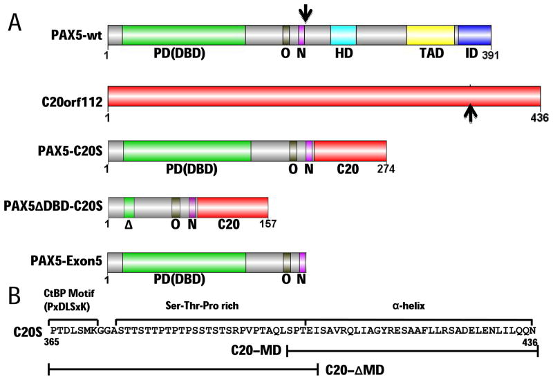
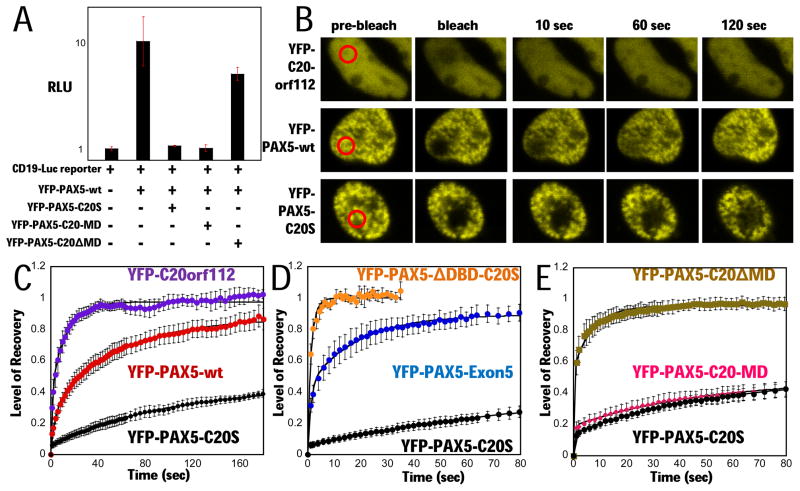
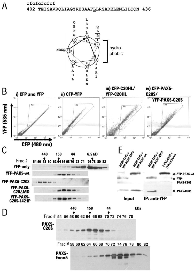
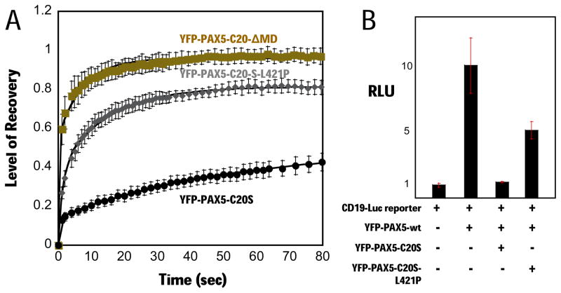
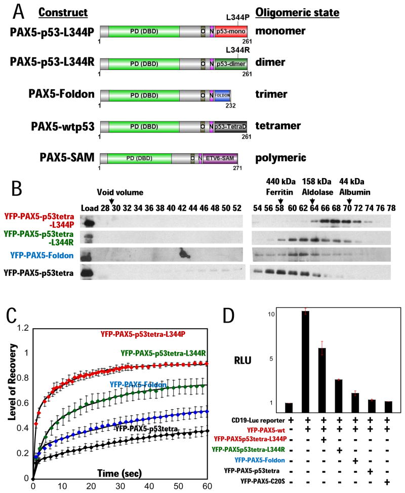
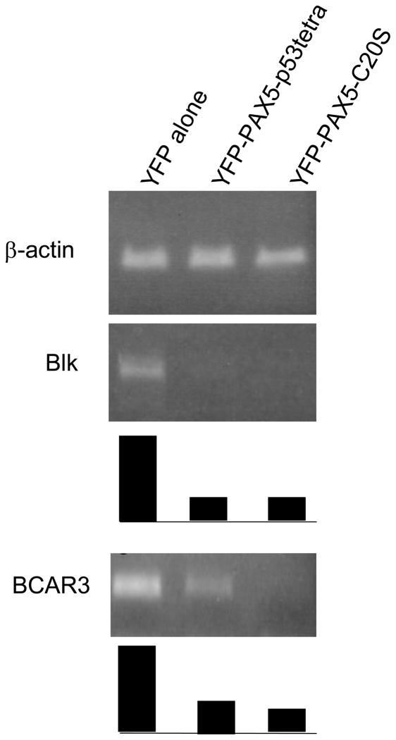
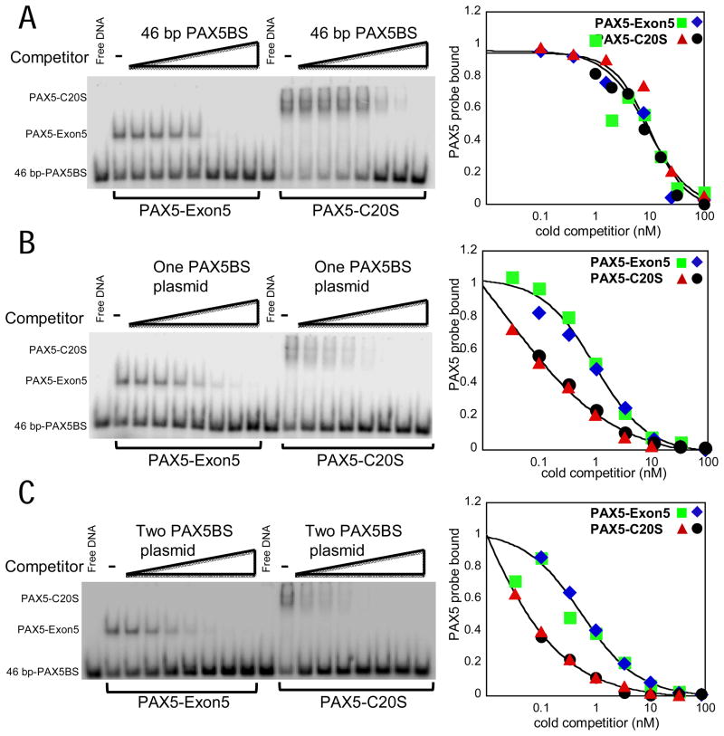
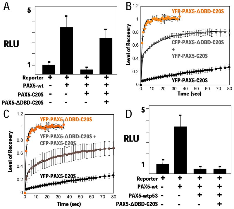
PAX5 encodes a master regulator of B-cell development. It fuses to other genes associated with acute lymphoblastoid leukemia (ALL). These fusion products are potent dominant-negative (DN) inhibitors of wild-type PAX5, resulting in a blockade of B-cell differentiation. Here, we show that multimerization of PAX5 DNA-binding domain (DBD) is necessary and sufficient to cause extremely stable chromatin binding and DN activity. ALL-associated PAX5-C20S results from fusion of the N-terminal region of PAX5, including its paired DBD, to the C-terminus of C20orf112, a protein of unknown function. We report that PAX5-C20S is a tetramer, which interacts extraordinarily stably with chromatin as determined by Fluorescence Recovery After Photobleaching in living cells. Tetramerization, stable chromatin binding and DN activity all require a putative five-turn amphipathic α-helix at the C-terminus of C20orf112, and does not require potential corepressor binding peptides elsewhere in the sequence. In vitro, the monomeric PAX5 DBD and PAX5-C20S binds a PAX5-binding site with equal affinity when it is at the center of an oligonucleotide too short to bind to more than one PAX5 DBD. But, PAX5-C20S binds the same sequence with 10-fold higher affinity than the monomeric PAX5 DBD when it is in a long DNA molecule. We suggest that the increased affinity results from interactions of one or more of the additional DBDs with neighboring non-specific sites in a long DNA molecule, and that this can account for the increased stability of PAX5-C20S chromatin binding compared with wild-type PAX5, resulting in DN activity by competition for binding to PAX5-target sites. Consistent with this model, the ALL-associated PAX5 fused to ETV6 or the multimerization domain of ETV6 SAM results in stable chromatin binding and DN activity. In addition, PAX5 DBD fused to artificial dimerization, trimerization and tetramerization domains results in parallel increases in the stability of chromatin binding and DN activity. Our studies suggest that oncogenic fusion proteins that retain the DBD of the transcription factor (TF) and the multimerization sequence of the partner protein can act in a DN manner by multimerizing and binding avidly to gene targets, preventing the normal TF from binding and inducing expression of its target genes. Inhibition of this multimeriztion may provide a novel therapeutic approach for cancers with this or similar fusion proteins.


Figures

Figure 1
Figure 1. Structural determinants of PAX5-C20orf112 oncogenic fusion protein
(A) Domains of PAX5 (Cobaleda et al., 2007), C20orf112, PAX5-C20S, PAX5ΔDBD-C20S with the paired DBD deleted, and PAX5-Exon5 (encoded by PAX5 exons 1–5, the portion of PAX5 in PAX5-C20S). The PAX5 DNA-binding domain [PD (DBD)], an octapeptide motif bound by Groucho-family co-repressors (O), and a nuclear localization sequence (N) are included in PAX5-C20S while its partial homeodomain (HD), activation domain (TAD), and inhibitory domain (ID) are not. Black arrows indicate points of fusion. (B) Analysis of C20orf112 sequence in PAX5-C20S reveals three regions that may contribute to DN activity: a CtBP binding motif (P×DLS×K), a serine-threonine-proline (Ser-Thr-Pro)-rich region, and a putative α-helical region. Constructs that contain bracketed parts of C20S are YFP-PAX5-C20-MD that fuses only the α-helical region, and YFP-PAX5-C20ΔMD that fuses only the P×DLS×K motif and the Ser-Thr-Pro rich region.
Figure 2
Figure 2. PAX5-C20S suppresses wt PAX5 activity and causes extremely slow nuclear mobility in living cells
(A) A YFP-PAX5-wt vector and the luciferase reporter were either transfected or co-transfected with vectors for YFP-PAX5-C20S, YFP-PAX5-C20-MD, or YFP-PAX5-C20ΔMD and luciferase assayed (RLU). (B) Fluorescence micrographs from FRAP assays in 293T cells. Red circled areas were photobleached regions, and fluorescence signal in each area was sequentially measured. FRAP assays of (C) YFP-PAX5-C20S (black circles, N=4, t1/2 ~ 200 sec), YFP-C20orf112 (purple circles, N=4, t1/2 ~ 3 s) and YFP-PAX5-wt (red circles, N=7, t1/2 ~ 24 s) in 293T cells, (D) YFP-PAX5-ΔDBD-C20S (orange circles, N=3, t1/2 ~ 1 s), YFP-PAX5-C20S (black circles, N=4, t1/2 ~ 200 s), and YFP-PAX5-Exon5 (blue circles, N=3, t1/2 ~ 4 s) in 293T cells, (E) YFP-PAX5-C20ΔMD (gold squares, N=6, t1/2 ~ 1 s), YFP-PAX5-C20-MD (pink diamonds, N= 7, t1/2 ~200 s), and YFP-PAX5-C20S (black circles, N=5, t1/2 ~200 s) in H1299 cells. Error bars here and in subsequent FRAP assays represent standard error of the mean.
Figure 3
Figure 3. α-helical region of C20S mediates multimerization
(A) Helical wheel projection of the predicted α-helix (aa 402–436). Heptad positions are labeled by letters ‘a’ through ‘g’ in the wheels. Hydrophobic residues occur at positions ‘c’ and ‘f’. The leucine at position 421 mutated to proline is boxed in projection and underlined in the sequence (B) Results of FRET assays. CFP or YFP fusion proteins were transiently expressed in 293T cells, and FRET signals were detected by flow cytometry. Cells were excited with a 436 nm laser, and emission signals were measured at 480 nm (x axis) and 535 nm (y axis). When FRET occurs, yellow signals (535 nm, y axis) increase. Signals in the triangular areas indicate cells with FRET signals. In a negative control (i), independent CFP and YFP proteins were co-expressed and produced no significant FRET signal (rare signals in the triangular area). A YFP-CFP fusion protein was expressed and used as a positive control (ii), showing that most cells exhibited strong FRET signals. (iii) Expression of YFP-C20HL and CFP-C20HL produced FRET signals comparable to the YFP-CFP fusion positive control. (iv) Expression of YFP-PAX5-C20S and CFP-PAX5-C20S produced a FRET signal in most cells of lower magnitude because of the greater distance between the YFP and CFP. (C) Gel filtration analysis of YFP, YFP-PAX5, YFP-PAX5-C20S, YFP-PAX5-C20-ΔMD, and YFP-PAX5-C20S-L421P. Proteins were expressed in H1299 cells, extracted from nuclei, and fractionated by gel filtration. Column fractions were subjected to immunoblotting with anti-YFP antibody. Fraction numbers are indicated above panel with protein standards eluted at specific volumes as indicated by arrows. (D) Gel filtration of PAX5-C20S-His6 and PAX5-Exon5-His6 expressed in and purified from E. coli. (E) Co-immunoprecipitation assays. (left panel: Input) PAX5-C20S was co-expressed with either YFP-PAX5-C20S or YFP-PAX5-wt in 293T cells. Expression of each protein was confirmed by immunoblotting cell extracts with YFP-specific antibody (top panel) or antibody specific for the C20 C-terminus (bottom panel). (Right panel) extracts were immunoprecipitated using YFP-antibody and immunoblotted as for the input panels.
Figure 4
Figure 4. Disruption of C20S multimerization abrogates suppression of PAX5 activity and stable chromatin binding
(A) FRAP assay comparing YFP-PAX5-C20-ΔMD (gold squares, N=5, t1/2 ~ 2 s), YFP-PAX5-C20S-L421P (gray diamonds, N=5, t1/2 ~ 5 s), and YFP-PAX5-C20S (black circles, N=4, t1/2 ~ 200 s) at 24 h post-transfection in H1299 cells. (B) YFP-PAX5-wt and the luciferase reporter were co-transfected alone, or with YFP-PAX5-C20S, YFP-PAX5-C20-MD, YFP-PAX5-C20-ΔMD, and YFP-PAX5-C20S-L421P, and luciferase activity assayed.
Figure 5
Figure 5. Multimerization of PAX5 fusions mediates suppression of wt PAX5 activity and stable chromatin binding
(A) Structure of PAX5-fusions: PAX5-p53tetra-L344P (monomer), PAX5-p53tetra-L344R (dimer), PAX5-Foldon (trimer), and PAX5-p53tetra (tetramer) domain. The fusions include the same N-terminal region of PAX5 in PAX5-C20S fused to the oligomerization domain of the fusion partner, p53 (aa 305–360) and foldon (aa 458–484). PAX5-SAM fusion contains the same region of PAX5 fused to the ETV6 SAM multimerization domain. (B) Gel filtration analysis of YFP-PAX5-fusions. Proteins were expressed in H1299 cells, extracted, fractionated by gel filtration, and detected by immunoblotting with anti-YFP antibody. (C) FRAP assays with YFP-PAX5 fusions in H1299 cells. FRAP recovery curves of YFP-PAX5-L344P (red diamonds, N= 3, t1/2 ~ 3 s), YFP-PAX5-L344R (green diamonds, N=3, t1/2 ~ 14 s), YFP-PAX5-Foldon (blue diamonds, N=5, t1/2 ~ 50 s), and YFP-PAX5-wtp53tetra (black diamonds, N=5, t1/2 ~ 200 s) show that increasing multimerization of the PAX5 DNA binding domain results in increasing stability of chromatin association. (D) YFP-PAX5wt and reporter gene were co-transfected either alone or with YFP-PAX5-L344P, YFP-PAX5-L344R, YFP-PAX5-Foldon, YFP-PAX5-wtp53tetra, or YFP-PAX5-C20S in p53-minus H1299 cells. Increasing multimerization of the PAX5 DNA binding domain caused increasing suppression of YFP-PAX5wt activity. (E) Semi-quantitative RT-PCR. Expression levels of two major PAX5 downstream target genes, Blk and BCAR3, as well as beta-actin (internal control) were examined by semi-quantitative RT-PCR. Products were amplified from cDNA of Burkit B-cell line, Daudi transfected with either YFP, YFP-PAX5-p53tetra, or YFP-PAX5-C20S (PCR cycle for Beta-actin: 25; PCR cycles for Blk: 30; PCR cycles for BCAR3: 33). Ten microliter of each PCR product was run in 2% agarose gel. Relative semi-quantitative levels of each PCR product to beta-actin PCR product are graphed uner each panel of PCR bands; levels of PCR products from Daudi cells transfected with YFP alone were regarded as 1.
Figure 6
Figure 6. PAX5-C20S tetramer has greater affinity in vitro for a PAX5 site in a long DNA molecule than monomeric PAX5-Exon5
(A) EMSA using a 46 bp 32P-labeled high affinity PAX5 binding site (PAX5BS) derived from the CD19 promoter (Czerny et al., 1993). Increasing amounts of unlabeled 46-bp PAX5BS (0.1, 0.4, 1.6, 6.4, 26, 100, 400 nM) were added before addition of protein to the binding reaction. The fraction of labeled 46-bp PAX5BS probe bound by protein (PAX5-Exon5 or PAX5-C20S) was calculated and plotted to the right relative to the amount bound without competitor. Results from two independent assays are indicated by the green squares and blue diamond symbols for PAX5-Exon5 and the red triangles and black circles for PAX5-C20. (B, C) Performed as in (A) except the unlabeled competitor DNA (0.1, 0.3, 1.0, 3, 10, 30, 100 nM) was a 5.15 kb plasmid with one or two copies (separated by 2 kb) of the 46 bp PAX5BS cloned into it. The data were fit by the variable slope sigmoid equation (see Experimental Procedures, Supplemental Materials).
Figure 7
Figure 7. The C20S tetramerization domain suppresses the dominant negative activity of PAX5-C20S
(A) Vectors for PAX5-wt, PAX5-C20S, and PAX5-C20SΔDBD and the luciferase reporter were transfected into 293T cells as indicated and luciferase assayed after 24 h. FRAP assays for (B) YFP-PAX5-ΔDBD-C20S (orange circles, N=3, t1/2 ~ 1 s), YFP-PAX5-C20S co-expressed with four-fold more CFP-PAX5-ΔDBD-C20S (YFP FRAP gray diamonds, N=7, t1/2 ~ 4 s), and YFP-PAX5-C20S (black circles, N=4, t1/2 ~ 200 s), and for (C) YFP-PAX5-ΔDBD-C20S (orange circles, N=3, t1/2 ~ 1 s), YFP-PAX5-ΔDBD-C20S and an equal amount of CFP-PAX5-C20S (gray circles, N=8, t1/2 ~ 25 s), and YFP-PAX5-C20S (black circles, N=4, t1/2 ~ 200 s). (D) PAX5-wtp53tetra was expressed with wt PAX5 and PAX5 reporter gene either with or without PAX5-C20SΔDBD and luciferase assayed.

Similar articles

Cited by

References

    1. An Q, Wright SL, Konn ZJ, Matheson E, Minto L, Moorman AV, et al. Variable breakpoints target PAX5 in patients with dicentric chromosomes: a model for the basis of unbalanced translocations in cancer. Proc Natl Acad Sci U S A. 2008;105:17050–4. - PMC - PubMed
    1. Antolini F, Lo Bello M, Sette M. Purified promyelocytic leukemia coiled-coil aggregates as a tetramer displaying low alpha-helical content. Protein Expr Purif. 2003;29:94–102. - PubMed
    1. Bousquet M, Broccardo C, Quelen C, Meggetto F, Kuhlein E, Delsol G, et al. A novel PAX5-ELN fusion protein identified in B-cell acute lymphoblastic leukemia acts as a dominant negative on wild-type PAX5. Blood. 2007;109:3417–23. - PubMed
    1. Cazzaniga G, Daniotti M, Tosi S, Giudici G, Aloisi A, Pogliani E, et al. The paired box domain gene PAX5 is fused to ETV6/TEL in an acute lymphoblastic leukemia case. Cancer Res. 2001;61:4666–70. - PubMed
    1. Cobaleda C, Schebesta A, Delogu A, Busslinger M. Pax5: the guardian of B cell identity and function. Nat Immunol. 2007;8:463–70. - PubMed

Publication types

MeSH terms