. 2011;6(7):e21891.
doi: 10.1371/journal.pone.0021891. Epub 2011 Jul 12.

Gastrodin inhibits expression of inducible NO synthase, cyclooxygenase-2 and proinflammatory cytokines in cultured LPS-stimulated microglia via MAPK pathways

Affiliations

Gastrodin inhibits expression of inducible NO synthase, cyclooxygenase-2 and proinflammatory cytokines in cultured LPS-stimulated microglia via MAPK pathways

Ji-Nan Dai et al. PLoS One. 2011.

Abstract

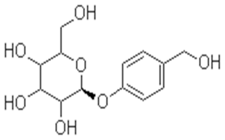
Background: Microglial activation plays an important role in neurodegenerative diseases by producing several proinflammatory enzymes and proinflammatory cytokines. The phenolic glucoside gastrodin, a main constituent of a Chinese herbal medicine, has been known to display anti-inflammatory properties. The current study investigates the potential mechanisms whereby gastrodin affects the expression of potentially pro-inflammatory proteins by cultured murine microglial BV-2 cells stimulated with lipopolysaccharide (LPS).

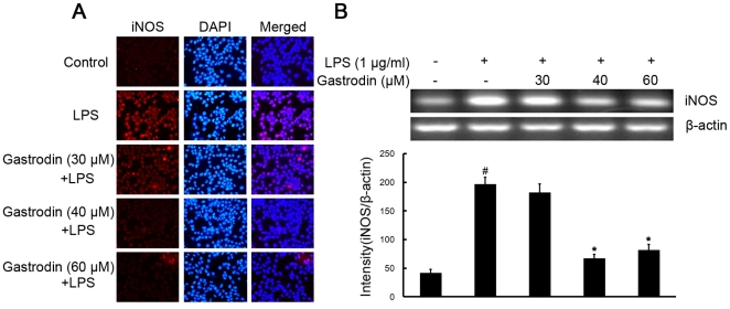
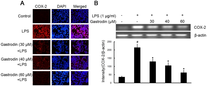
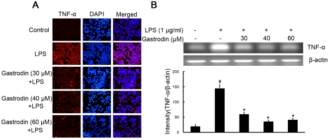
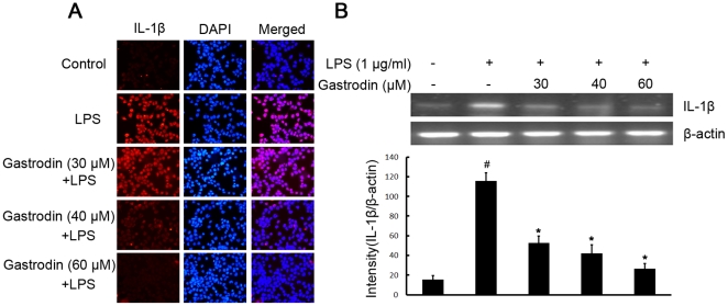
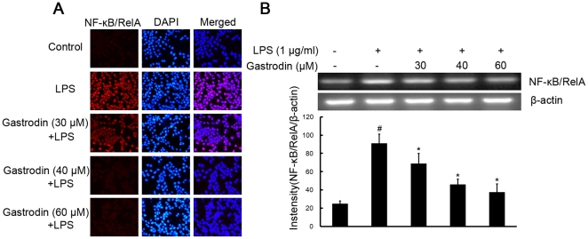
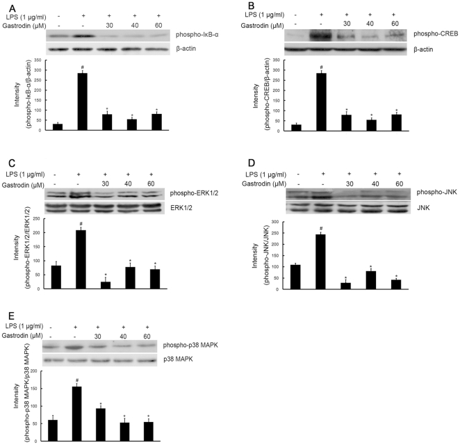
Methodology/principal findings: BV-2 cells were pretreated with gastrodin (30, 40, and 60 µM) for 1 h and then stimulated with LPS (1 µg/ml) for another 4 h. The effects on proinflammatory enzymes, inducible nitric oxide synthase (iNOS) and cyclooxygenase-2 (COX-2), and proinflammatory cytokines, tumor necrosis factor-α (TNF-α), and interleukin-1β (IL-1β), are analysed by double-immunofluorescence labeling and RT-PCR assay. To reveal the mechanisms of action of gastrodin we investigated the involvement of mitogen-activated protein kinases (MAPKs) cascades and their downstream transcription factors, nuclear factor-κB (NF-κB) and cyclic AMP-responsive element (CRE)-binding protein (CREB). Gastrodin significantly reduced the LPS-induced protein and mRNA expression levels of iNOS, COX-2, TNF-α, IL-1β and NF-κB. LPS (1 µg/ml, 30 min)-induced phosphorylation of extracellular signal-regulated kinase 1/2 (ERK1/2), c-Jun N-terminal protein kinase (JNK) and p38 mitogen-activated protein kinase (p38 MAPK) and this was inhibited by pretreatment of BV-2 cells with different concentrations of gastrodin (30, 40, and 60 µM). In addition, gastrodin blocked LPS-induced phosphorylation of inhibitor κB-α (IκB-α) (and hence the activation of NF-κB) and of CREB, respectively.

Conclusion and implications: This study indicates that gastrodin significantly attenuate levels of neurotoxic proinflammatory mediators and proinflammatory cytokines by inhibition of the NF-κB signaling pathway and phosphorylation of MAPKs in LPS-stimulated microglial cells. Arising from the above, we suggest that gastrodin has a potential as an anti-inflammatory drug candidate in neurodegenerative diseases.


Conflict of interest statement

Competing Interests: One of the authors, Y. D. Liu, is employed by a commercial company, Kunming Pharmaceutical Corporation. This does not alter the authors' adherence to all the PLoS ONE policies on sharing data and materials.

Figures

Figure 1
Figure 1. Structure of gastrodin.
Figure 2
Figure 2. Inhibitory effects of gastrodin on the LPS-induced expression of protein and mRNA for iNOS in BV-2 cells.
Approximately 1×10⁶ cells/ml were seeded in six-well plates and incubated until 80% confluency. Cells were pre-treated with gastrodin (30, 40, and 60 µM) for 1 h, then exposed to 1 µg/ml LPS for 4 h. The levels of protein and the corresponding mRNA were determined by double-immunofluorescence labeling and RT-PCR as described in the Methods. Panel A shows the immunofluorenscence images for protein expression of iNOS and Panel B shows the corresponding mRNA data. The relative mRNA level was quantified by scanning densitometry and normalized to β-actin mRNA. The values shown are mean ± SEM of data from three independent experiments. # Significant compared with control alone, p<0.05. *Significant compared with LPS alone, p<0.05.
Figure 3
Figure 3. Inhibitory effects of gastrodin on the LPS-induced expression of protein and mRNA for COX-2 in BV-2 cells.
Approximately 1×10⁶ cells/ml were seeded in six-well plates and incubated until 80% confluency. Cells were pre-treated with gastrodin (30, 40, and 60 µM) for 1 h, then exposed to 1 µg/ml LPS for 4 h. The levels of protein and the corresponding mRNA were determined by double-immunofluorescence labeling and RT-PCR as described in the Methods. Panel A shows the immunofluorenscence images for protein expression of COX-2 and Panel B shows the corresponding mRNA data. The relative mRNA level was quantified by scanning densitometry and normalized to β-actin mRNA. The values shown are mean ± SEM of data from three independent experiments. # Significant compared with control alone, p<0.05. *Significant compared with LPS alone, p<0.05.
Figure 4
Figure 4. Inhibitory effects of gastrodin on the LPS-induced expression of protein and mRNA for TNF-α in BV-2 cells.
Approximately 1×10⁶ cells/ml were seeded in six-well plates and incubated until 80% confluency. Cells were pre-treated with gastrodin (30, 40, and 60 µM) for 1 h, then exposed to 1 µg/ml LPS for 4 h. The levels of protein and the corresponding mRNA were determined by double-immunofluorescence labeling and RT-PCR as described in the Methods. Panel A shows the immunofluorenscence images for protein expression of TNF-α and Panel B shows the corresponding mRNA data. The relative mRNA level was quantified by scanning densitometry and normalized to β-actin mRNA. The values shown are mean ± SEM of data from three independent experiments. # Significant compared with control alone, p<0.05. *Significant compared with LPS alone, p<0.05.
Figure 5
Figure 5. Inhibitory effects of gastrodin on the LPS-induced expression of protein and mRNA for IL-1β in BV-2 cells.
Approximately 1×10⁶ cells/ml were seeded in six-well plates and incubated until 80% confluency. Cells were pre-treated with gastrodin (30, 40, and 60 µM) for 1 h, then exposed to 1 µg/ml LPS for 4 h. The levels of protein and the corresponding mRNA were determined by double-immunofluorescence labeling and RT-PCR as described in the Methods. Panel A shows the immunofluorenscence images for protein expression of IL-1β and Panel B shows the corresponding mRNA data. The relative mRNA level was quantified by scanning densitometry and normalized to β-actin mRNA. The values shown are mean ± SEM of data from three independent experiments. # Significant compared with control alone, p<0.05. *Significant compared with LPS alone, p<0.05.
Figure 6
Figure 6. Inhibitory effects of gastrodin on the LPS-induced expression of protein and mRNA for NF-κB/RelA in BV-2 cells.
Approximately 1×10⁶ cells/ml were seeded in six-well plates and incubated until 80% confluency. Cells were pre-treated with gastrodin (30, 40, and 60 µM) for 1 h, then exposed to 1 µg/ml LPS for 4 h. The levels of protein and the corresponding mRNA were determined by double-immunofluorescence labeling and RT-PCR as described in the Methods. Panel A shows the immunofluorenscence images for protein expression of NF-κB/RelA and Panel B shows the corresponding mRNA data. The relative mRNA level was quantified by scanning densitometry and normalized to β-actin mRNA. The values shown are mean ± SEM of data from three independent experiments. # Significant compared with control alone, p<0.05. *Significant compared with LPS alone, p<0.05.
Figure 7
Figure 7. Inhibitory effects of gastrodin on the LPS-induced phosphorylation expression of IκB-α(A), CREB (B), ERK1/2 (C), JNK(D) and p38 MAPK (E) in BV-2 cells.
Approximately 1×10⁶ cells/ml were seeded in six-well plates and incubated until 80% confluency. Cells were pre-treated with gastrodin (30, 40, and 60 µM) for 1 h, then exposed to 1 µg/ml LPS for 30 min. Cell lysates (50 µg protein) were prepared and subjected to Western blot analysis by using antibodies specific for phosphorylated forms of IκB-α, CREB, ERK1/2, JNK and p38 MAPK (shown as phospho-IκB-α, etc.) as described in the methods. Equivalent loading of cell lysates was determined by reprobing the blots with anti-β-actin, total ERK1/2, JNK or p38 MAPK antibodies. The relative protein levels were quantified by scanning densitometry and normalized to β-actin, total ERK1/2, JNK or p38 MAPK. The values shown are mean ± SEM of data from three independent experiments. # Significant compared with control alone, p<0.05. *Significant compared with LPS alone, p<0.05.

References

    1. Olson JK, Miller SD. Microglia initiate central nervous system innate and adaptive immune responses through multiple TLRs. J Immunol. 2004;173:3916–3924. - PubMed
    1. Nakajima K, Kohsaka S. Functional roles of microglia in the central nervous system. Hum Cell. 1998;11:141–155. - PubMed
    1. Suzumura A, Takeuchi H, Zhang G, Kuno R, Mizuno T. Roles of glia-derived cytokines on neuronal degeneration and regeneration. Ann N Y Acad Sci. 2006;1088:219–229. - PubMed
    1. Roy A, Fung YK, Liu X, Pahan K. Up-regulation of Microglial CD11b Expression by Nitric Oxide. J Biol Chem. 2006;281:14971–14980. - PMC - PubMed
    1. Ankeny DP, Popovich PG. Mechanisms and implications of adaptive immune responses after traumatic spinal cord injury. Neuroscience. 2009;158:1112–1121. - PMC - PubMed

Publication types

MeSH terms