. 2011;6(7):e21922.
doi: 10.1371/journal.pone.0021922. Epub 2011 Jul 13.

Extension of lifespan in C. elegans by naphthoquinones that act through stress hormesis mechanisms

Affiliations

Extension of lifespan in C. elegans by naphthoquinones that act through stress hormesis mechanisms

Piper R Hunt et al. PLoS One. 2011.

Abstract

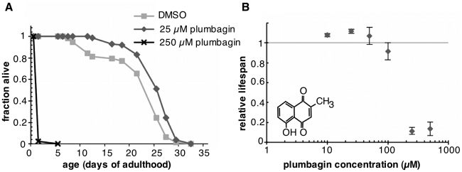
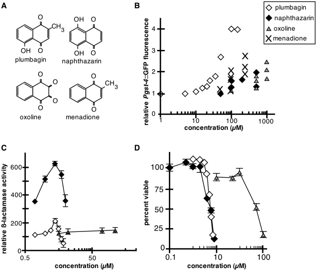
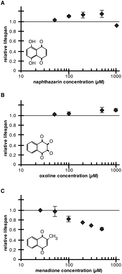
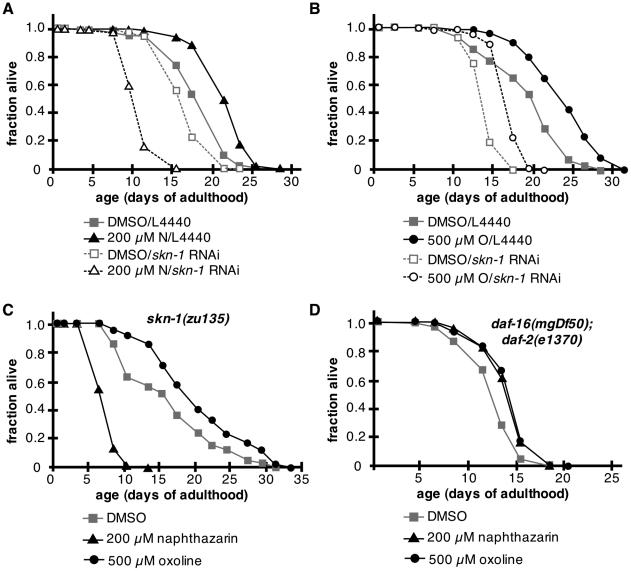
Hormesis occurs when a low level stress elicits adaptive beneficial responses that protect against subsequent exposure to severe stress. Recent findings suggest that mild oxidative and thermal stress can extend lifespan by hormetic mechanisms. Here we show that the botanical pesticide plumbagin, while toxic to C. elegans nematodes at high doses, extends lifespan at low doses. Because plumbagin is a naphthoquinone that can generate free radicals in vivo, we investigated whether it extends lifespan by activating an adaptive cellular stress response pathway. The C. elegans cap'n'collar (CNC) transcription factor, SKN-1, mediates protective responses to oxidative stress. Genetic analysis showed that skn-1 activity is required for lifespan extension by low-dose plumbagin in C. elegans. Further screening of a series of plumbagin analogs identified three additional naphthoquinones that could induce SKN-1 targets in C. elegans. Naphthazarin showed skn-1dependent lifespan extension, over an extended dose range compared to plumbagin, while the other naphthoquinones, oxoline and menadione, had differing effects on C. elegans survival and failed to activate ARE reporter expression in cultured mammalian cells. Our findings reveal the potential for low doses of naturally occurring naphthoquinones to extend lifespan by engaging a specific adaptive cellular stress response pathway.


Conflict of interest statement

Competing Interests: The authors have declared that no competing interests exist.

Figures

Figure 1
Figure 1. Low-dose plumbagin increases mean lifespan in C. elegans by stress hormesis.
(A) Survival of wildtype adults treated with DMSO vehicle, 25 or 250 µM plumbagin. Mean lifespan was extended by 25 µM plumbagin (n = 137, p<0.0001) while the 250 µM dose was toxic and shortened lifespan (n = 144, p<0.0001). (B) Dose-response curve for mean survival relative to plumbagin concentration (µM). The plot shows an inverted U-shaped curve characteristic of stress hormesis. Error bars indicate variation in two to four independent lifespan experiments per dose. Individual trial data are presented in Table S1.
Figure 2
Figure 2. Transcriptional effects of plumbagin under non-toxic conditions in C. elegans.
(A,B) Results of GO analysis of molecular function (A) and biological processes (B) for transcripts differently expressed in animals treated with plumbagin versus vehicle (DMSO). Results indicate upregulation of genes involved in oxidative stress resistance functions (A) and biological functions related electron transport (B), possible related to electron capture. Furthermore, reductions in growth and developmental processes were identified in plumbagin-treated animals, consistent with a shift of resources to stress response.
Figure 3
Figure 3. Effect of plumbagin on adult lifespan of skn-1 and daf-16 mutants.
(A–C) Mean adult lifespan on 25 µM plumbagin (filled columns) compared to vehicle control (unfilled columns) was measured in 4 independently-conducted trials for (A) wildtype, (B) skn-1(zu135) and (C) daf-16(mgDf50); daf-2(e1370) adults. Asterisks mark trails in which mean adult lifespan was extended by 25 µM plumbagin treatment with Log-Rank probability > 0.01. Lifespan data for additional plumbagin doses is presented on Table S1. (D) Dose-response curve of daf-16(mgDf50) adult lifespan at 0, 10, 25, 50 and 100 µM plumbagin. Asterisk indicates a positive effect of plumbagin on lifespan with Log-Rank probability > 0.01. Data for lifespan trials is presented in Table S1.
Figure 4
Figure 4. Effect of low-dose plumbagin on DAF-16, SKN-1 and ARE transcriptional reporters in C. elegans and HepG2 cells.
(A) Dose-response curve for Pgst-4::GFP fluorescence versus plumbagin concentration. Levels of Pgst-4::GFP were determined for a minimum of 20 worms per condition per trial by average background-subtracted values for whole worm fluorescence of treated animals normalized to controls; p<0.0001 (T-test) in every trial at doses of 20 µM plumbagin and above. Each data point represents a single trial. Results for individual trials are shown in Table S2. (B) Representative images of Pgst-4::GFP in wildtype day 1 adults treated for two days with 25 µM plumbagin under control conditions (left panel) or with skn-1 RNAi (right panel). In control animals, 25 µM plumbagin was associated with increased Pgst-4::GFP in the intestine and muscles and skn-1 RNAi abrogated the increases in Pgst-4::GFP fluorescence levels. Bar, 0.2 mm. (C) ARE reporter activity in HepG2 cells treated with DMSO vehicle control (open) or 4 µM plumbagin (shaded). Plumbagin activated ARE reporter beta-lactamase expression. Co-incubation with reduced glutathione significantly reduced induction of the ARE reporter by plumbagin. Data are relative beta-lactamase activity + SEM. (D) Adult TH356 hermaphrodites expressing a DAF-16::GFP translational fusion were treated as indicated and localization of DAF-16::GFP monitored as a measure of DAF-16 activation. Thermal stress (37°C, 20 minutes) induced dramatic nuclear accumulation of DAF-16::GFP in intestinal nuclei. In contrast, DMSO vehicle and 25 µM plumbagin treatments failed to induce nuclear DAF-16::GFP accumulation. Two independent experiments were performed with n = 20 animals/treatment. For DMSO and plumbagin, animals were transferred to treatments and DAF-16:GFP localization was scored after 3, 24 and 48 hours. Shown are representative 48-hour treatments. Bar, 0.1 mm; all images were collected using identical exposure times.
Figure 5
Figure 5. Effects of naphthazarin, oxoline and menadione on skn-1 and Nrf2 targets in C. elegans and HepG2 cells.
(A) Structures of plumbagin, naphthazarin, oxoline and menadione. (B) Induction of Pgst-4::GFP by naphthazarin, oxoline, and menadione. Data are results from independent experiments measuring whole worm Pgst-4::GFP fluorescence compared to vehicle-treated controls (Table S3). For comparison, the average level of Pgst-4::GFP fluorescence in plumbagin-treated animals is shown on the same plot (diamonds, data shown on Fig. 4A). Shaded diamonds, naphthazarin, n≥30, p<0.0001 for 100 µM and above; triangles, oxoline, n≥16, p<0.0001 for 500 µM and above; crosses, menadione, n≥18, p<0.0001 for 100 µM and above. (C) Plumbagin (diamonds) and naphthazarin (shaded diamonds) activated ARE reporter expression (beta-lactamase) in HepG2 cells. Toxicity at higher concentrations of plumbagin and naphthazarin resulted in decreased reporter gene expression. Oxoline (triangles) failed to induce expression of ARE driven beta-lactamase. The ARE reporter cell line HepG2 was exposed to the indicated concentrations of plumbagin, naphthazarin or oxoline and ARE-driven beta-lactamase activity was assessed 24 hrs after exposure. Data are mean beta-lactamase activity ± SEM (n = 3−4). (D) HepG2 cells were treated with plumbagin (diamonds), naphthazarin (shaded diamonds) or oxoline (triangles) at indicated concentrations. Cell viability was assessed 24 hrs after exposure. Results are the mean from 3−4 experiments +/− SEM.
Figure 6
Figure 6. Effects of naphthazarin, oxoline, and menadione on C. elegans survival.
(A–C) Dose-response relationship of relative C. elegans survival with respect to naphthoquinone concentration. Chemical structures for each compound are shown on each plot. (A) Naphthazarin increased C. elegans lifespan at higher doses than plumbagin. Data represent one trial at 50 µM (n = 67, p = 0.2), and three trials each at 100 µM (n≥93, p<0.0001 in 2 of 3 trials), 200 µM (n≥96, p<0.0001 in 3 of 3 trials), and 500 µM (n≥66, p<0.0001 in 2 of 3 trials). (B) Oxoline treatment extended mean lifespan at doses of 500 µM (n≥95, p<0.0001 in 3 of 4 trials) and 1 mM (n≥61, p<0.0001 in 2 of 2 trials). (C) Menadione treatment shortened C. elegans lifespan and did not provide a significant lifespan benefit at any tested dose (13 trials at various concentrations with n≥59 per condition, p<0.0001 in all trials at 100 µM and above).
Figure 7
Figure 7. Requirement for skn-1 in lifespan extension by naphthazarin.
(A) skn-1 RNAi abrogated lifespan extension by 200 µM naphthazarin. Filled squares, L4440 treated with DMSO vehicle control (n = 80); open squares, skn-1 RNAi treated with DMSO vehicle control (n = 125); filled triangles, L4440 treated with 200 µM naphthazarin (n = 88, p<0.0001); open triangles, skn-1 RNAi treated with 200 µM naphthazarin (n = 127, p<0.0001). (B) 500 µM oxoline was associated with similar increases in lifespan for both L4440 and skn-1 RNAi treated groups. Filled squares, L4440 treated with DMSO control (n = 117); open squares, skn-1 RNAi treated with DMSO vehicle control (n = 131); filled circles, L4440 treated with 500 µM oxoline (n = 106, p<0.0001); open circles, skn-1 RNAi treated with 500 µM oxoline (n = 124, p<0.0001). (C) Lifespan of skn-1(zu135) adults was shortened by 200 µM naphthazarin (triangles, n = 110, p<0.0001) relative to DMSO vehicle control (squares, n = 123), but lengthened by 500 µM oxoline (circles, n = 117, p<0.0001). (D) Mean lifespan of daf-16(mgDf50); daf-2(e1370) adults was extended by 200 µM naphthazarin (triangles, n = 116, p<0.0001) and 500 µM oxoline (circles, n = 121, p<0.0001), relative to DMSO vehicle treated controls (squares, n = 80).

References

    1. Finkel T, Holbrook NJ. Oxidants, oxidative stress and the biology of ageing. Nature. 2000;408:239–247. - PubMed
    1. Kregel KC, Zhang HJ. An integrated view of oxidative stress in aging: basic mechanisms, functional effects, and pathological considerations. Am J Physiol Regul Integr Comp Physiol. 2007;292:R18–36. - PubMed
    1. Bejarano E, Cuervo AM. Chaperone-mediated autophagy. Proc Am Thorac Soc. 2010;7:29–39. - PMC - PubMed
    1. Head E, Liu J, Hagen TM, Muggenburg BA, Milgram NW, et al. Oxidative damage increases with age in a canine model of human brain aging. J Neurochem. 2002;82:375–381. - PubMed
    1. Choksi KB, Nuss JE, Boylston WH, Rabek JP, Papaconstantinou J. Age-related increases in oxidatively damaged proteins of mouse kidney mitochondrial electron transport chain complexes. Free Radic Biol Med. 2007;43:1423–1438. - PMC - PubMed

Publication types

MeSH terms