Review
. 2011 Jun 1;15(3):145-58.
doi: 10.1007/BF03256405.

Molecular and nanotechnologic approaches to etiologic diagnosis of infectious syndromes

Affiliations
Review

Molecular and nanotechnologic approaches to etiologic diagnosis of infectious syndromes

Sathish Sankar et al. Mol Diagn Ther. .

Abstract

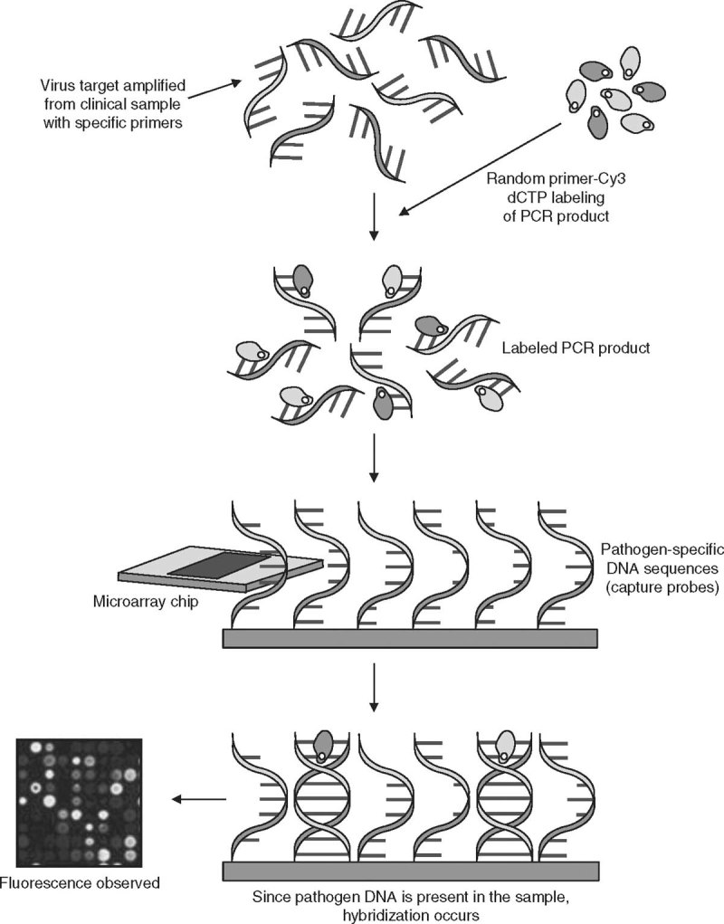
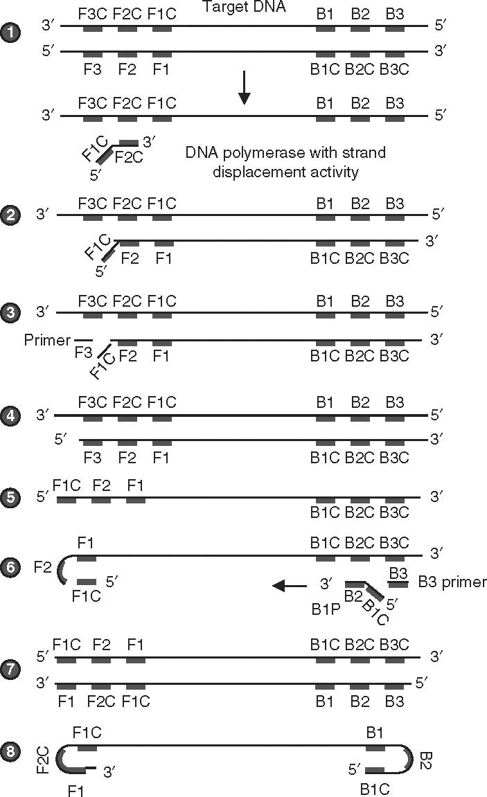
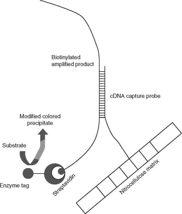
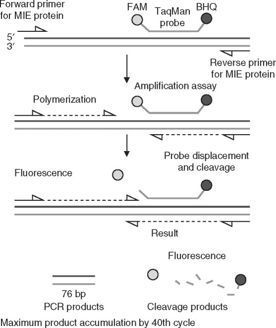
Infectious diseases are a major global public health problem. Multiple agents are now recognized to cause indistinguishable illnesses. The term 'syndrome' applies to such situations, for which early and rapid diagnosis of the infecting agent would enable prompt and appropriate therapy. Public health physicians would also get timely information on the specific etiology of the infectious syndrome, facilitating intervention at the community level in the face of outbreaks or epidemics. A variety of molecular techniques have been evaluated for rapid diagnosis of infectious syndromes. These techniques include real-time multiplex PCR, DNA microarray, loop-mediated isothermal amplification, and other similar assays. This review surveys such state-of-the-art technologies.


Figures

Fig. 1
Fig. 1
Principle of PCR-based DNA microarray detection of specific viral genes. A specific viral target gene is amplified from a clinical sample, using species-specific primers. The amplified PCR products are labeled with a random primer incorporated with Cy3 deoxycytidine triphosphate (dCTP). The labeled PCR products bind to the pathogen-specific oligonucleotide capture probe immobilized on the microarray chip. If pathogen DNA is present and amplified from the sample, hybridization with the capture probe is detected by analysis of fluorescence, using an Affymetrix 428 array scanner (Affymetrix, Santa Clara, CA, USA) with the aid of ImaGene software (version 4.2; BioDiscovery, Marina del Rey, CA, USA).
Fig. 2
Fig. 2
Sketch depicting the steps involved in the loop-mediated isothermal amplification (LAMP) assay. (1) The LAMP assay is entirely carried out at 65°C, when the double-stranded DNA (dsDNA) is in the state of dynamic equilibrium. In these conditions, one of the LAMP primers can anneal to the complimentary sequence of dsDNA (the target). Initiation of DNA synthesis is facilitated by DNA polymerase with strand displacement activity. The enzyme displaces and releases the single-stranded DNA (ssDNA). There is no denaturation step involved. The forward primers F1, F2, and F3 and the backward primers B1, B2, and B3 anneal to the complementary sequence (F1C, F2C, and F3C; B1C, B2C, and B3C) on one strand, extending the complementary strand sequence. Usually, six primers bind to about eight sites in the entire target sequence. (2) The DNA polymerase acts on the complementary DNA (cDNA) strand of the template DNA, starting from the 3′ end of the F2 region of the forward inner primer (FIP), and proceeding with strand synthesis. (3) The F3 primer anneals to the F3C region, releasing the FIP-linked complementary strand. (4) DNA synthesis proceeds from the F3 primer. (5) F1C (the FIP-linked complementary strand) is then released as a single strand because of displacement by the DNA strand synthesized from F3P, and the released single strand forms a stem-loop structure at the 5′ end. The loop formation occurs because F1C is complimentary to the F1 regions. (6) The ssDNA from step 5 serves as a template for backward inner primer (BIP)-initiated DNA synthesis. Subsequently, B3-primed strand displacement DNA synthesis occurs. The BIP anneals to the DNA strand produced in step 5. Starting from the 3′ end of the BIP, synthesis of the complementary strand takes place. This process allows the DNA to revert from a loop structure to a linear structure. The B3 primer anneals to the outside of the BIP and then, through the activity of DNA polymerase and starting at the 3′ end, the DNA synthesized from the BIP is displaced and released as a single strand before DNA synthesis from the B3 primer. (7) This process allows dsDNA synthesis. (8) After each amplification cycle, i.e. LAMP cycling, the BIP-linked complementary strand that is displaced forms a structure with a stem loop at each end (a dumbbell structure). The assays generally run for about 60 minutes, generating clear turbidity measured by a photometer. Usually, turbidity measured as absorbance of 0.1 is considered positive.
Fig. 3
Fig. 3
Schematic representation of the principle of the line probe assay. The nitrocellulose matrix (strip) carries pre-synthesized complementary DNA (cDNA) probes that are specific for genotypes of hepatitis C virus (HCV) immobilized at different locations; as many as 23 such probes, including amplification controls, are fixed on the matrix. The multiplex PCR product of HCV (produced with biotinylated primers) is applied in the reaction vessel containing the matrix. After hybridization, the captured amplicons are detected by a biotin-streptavidin enzyme conjugate with a substrate color reaction. The hybridization profile that is obtained is specific for each genotype, and this is interpreted with a standard chart.
Fig. 4
Fig. 4
Diagrammatic outline of the TaqMan-based real-time PCR assay for detection of cytomegalovirus. The fluorophore 6-carboxyfluorescein (FAM) is used as the probe label and Black Hole Quencher® (BHQ) is used as the quencher dye. Forward and reverse primers are targeted toward major immediate early (MIE) protein. Amplification is measured dynamically as fluorescence intensity. Usually, a cut-off fluorescence intensity of 0.05 or more with a typical sigmoid amplification curve within the 40th cycle is considered positive.
Fig. 5
Fig. 5
Graphic representation of the quantum dot (QD) principle. (a) QD nanocrystal used in analyte (microbial/viral antigen) detection. The QD (detection reagent) contains a core and shell component with the reagent, e.g. a monoclonal antibody specific for the analyte. (b) Optical principle underlying the detection of the analyte-reagent interaction on the surface of the QD, which results in a measurable variation that is detectable as a change in the photon emission pattern. QDs emit a narrow-band visible wavelength of light.

Similar articles

Cited by

References

    1. Tallury P, Malhotra A, Byrne LM, et al. Nanobioimaging and sensing of infectious diseases. Adv Drug Deliv Rev. 2010;62(4–5):424–37. doi: 10.1016/j.addr.2009.11.014. - DOI - PMC - PubMed
    1. Curr Opin Pediatr. 2009.
    1. Banér J, Gyarmati P, Yacoub A, et al. Microarray-based molecular detection of foot-and-mouth disease, vesicular stomatitis and swine vesicular disease viruses, using padlock probes. J Virol Methods. 2007;143(2):200–6. doi: 10.1016/j.jviromet.2007.03.004. - DOI - PubMed
    1. Hindson BJ, Reid SM, Baker BR, et al. Diagnostic evaluation of multiplexed reverse transcription-PCR microsphere array assay for detection of foot-and-mouth and look-alike disease viruses. J Clin Microbiol. 2008;46(3):1081–9. doi: 10.1128/JCM.01740-07. - DOI - PMC - PubMed
    1. Jack PJ, Amos-Ritchie RN, Reverter A, et al. Microarray-based detection of viruses causing vesicular or vesicular-like lesions in livestock animals. Vet Microbiol. 2009;133(1–2):145–53. doi: 10.1016/j.vetmic.2008.05.030. - DOI - PMC - PubMed

MeSH terms