. 2011 Jul 20;101(2):345-52.
doi: 10.1016/j.bpj.2011.05.058.

Defining the role of the tension sensor in the mechanosensitive channel of small conductance

Affiliations

Defining the role of the tension sensor in the mechanosensitive channel of small conductance

Hannah R Malcolm et al. Biophys J. .

Abstract

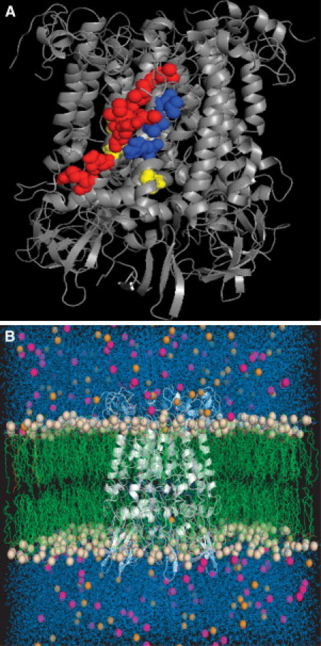
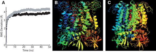
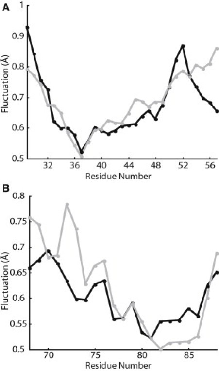
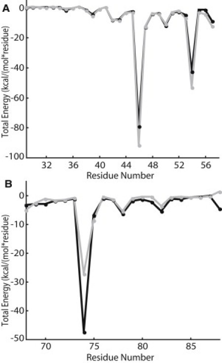
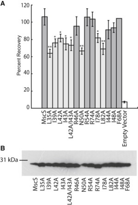
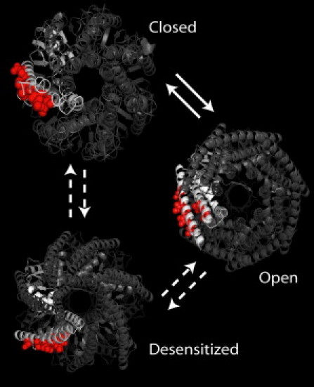
Mutations that alter the phenotypic behavior of the Escherichia coli mechanosensitive channel of small conductance (MscS) have been identified; however, most of these residues play critical roles in the transition between the closed and open states of the channel and are not directly involved in lipid interactions that transduce the tension response. In this study, we use molecular dynamic simulations to predict critical lipid interacting residues in the closed state of MscS. The physiological role of these residues was then investigated by performing osmotic downshock assays on MscS mutants where the lipid interacting residues were mutated to alanine. These experiments identified seven residues in the first and second transmembrane helices as lipid-sensing residues. The majority of these residues are hydrophobic amino acids located near the extracellular interface of the membrane. All of these residues interact strongly with the lipid bilayer in the closed state of MscS, but do not face the bilayer directly in structures associated with the open and desensitized states of the channel. Thus, the position of these residues relative to the lipid membrane appears related to the ability of the channel to sense tension in its different physiological states.


Figures

Figure 1
Figure 1
Closed state MscS model at the end of the MD simulation, as seen from the side (A) and with lipids, chloride ions, sodium ions, and water included in the simulation (B). The residues highlighted as a space-filling model in panel A correspond to (red) predicted lipid-interacting residues in TM1 (L35, I39, L42, I43, R46, N50, R54); (blue) predicted lipid interacting residues in TM2 (R74, I78, L82); and (yellow) noninteracting residues used as controls in this study (I44, I48, F68).
Figure 2
Figure 2
Structural deviations during the MD simulation. (A) RMS deviation of the MscS structure over the course of the MD simulation; Simulation 1 is shown in solid and Simulation 2 is shown in shaded representation. (B) Initial closed state all-atom MscS model generated with MODELLER. (C) MscS all-atom closed state model from Simulation 1 after 50 ns.
Figure 3
Figure 3
Average Cα RMS fluctuation per residue over the final 10 ns of the MD simulations for TM1 (A) and TM2 (B); Simulation 1 is shown in solid and Simulation 2 is shown in shaded representation.
Figure 4
Figure 4
Protein-lipid interactions energies averaged over the final 10 ns of the MD simulations for TM1 (A) and TM2 (B); Simulation 1 is shown in solid and Simulation 2 is shown in shaded representation.
Figure 5
Figure 5
Functional analysis of MscS alanine mutations. (A) Osmotic downshock assay results of wild-type MscS and alanine mutants. Error bars represent the standard error of the mean for six independent trials. Statistically partial loss of function mutations (light shaded bar) were determined by comparison with wild-type MscS using a Student's t-test (∗∗ = p < 0.01 and  = p < 0.05). (B) Western blot analysis of protein expression levels for wild-type MscS and alanine mutants under expression conditions identical to those used for the osmotic downshock assay.
Figure 6
Figure 6
Positions of the tension-sensing residues are shown in the closed (50-ns simulation), open (2VV5 (46)), and desensitized (2OAU (18,47)) states of MscS.

References

    1. Li C., Edwards M.D., Booth I.R. Identification of mutations that alter the gating of the Escherichia coli mechanosensitive channel protein, MscK. Mol. Microbiol. 2007;64:560–574. - PMC - PubMed
    1. Levina N., Tötemeyer S., Booth I.R. Protection of Escherichia coli cells against extreme turgor by activation of MscS and MscL mechanosensitive channels: identification of genes required for MscS activity. EMBO J. 1999;18:1730–1737. - PMC - PubMed
    1. Schumann U., Edwards M.D., Booth I.R. YbdG in Escherichia coli is a threshold-setting mechanosensitive channel with MscM activity. Proc. Natl. Acad. Sci. USA. 2010;107:12664–12669. - PMC - PubMed
    1. Sukharev S.I., Blount P., Kung C. A large-conductance mechanosensitive channel in E. coli encoded by MscL alone. Nature. 1994;368:265–268. - PubMed
    1. Hurst A.C., Petrov E., Martinac B. MscS, the bacterial mechanosensitive channel of small conductance. Int. J. Biochem. Cell Biol. 2008;40:581–585. - PubMed

Publication types

LinkOut - more resources