. 2011 Jul 20;101(2):411-20.
doi: 10.1016/j.bpj.2011.05.039.

Reengineering rate-limiting, millisecond enzyme motions by introduction of an unnatural amino acid

Affiliations

Reengineering rate-limiting, millisecond enzyme motions by introduction of an unnatural amino acid

Eric D Watt et al. Biophys J. .

Abstract

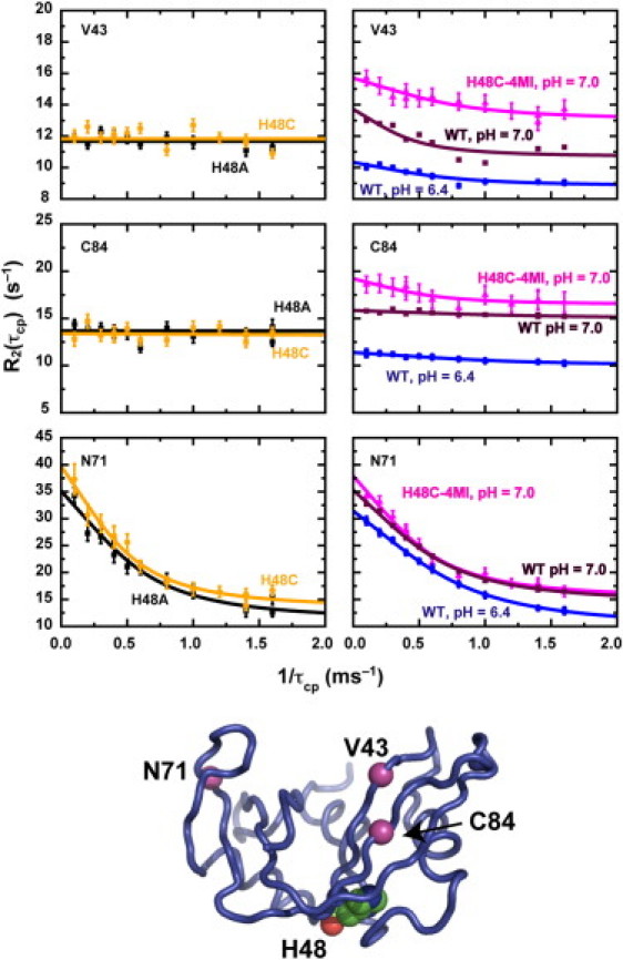
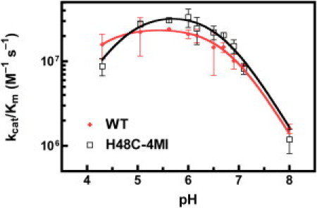
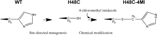
Rate-limiting millisecond motions in wild-type (WT) Ribonuclease A (RNase A) are modulated by histidine 48. Here, we incorporate an unnatural amino acid, thia-methylimidazole, at this site (H48C-4MI) to investigate the effects of a single residue on protein motions over multiple timescales and on enzyme catalytic turnover. Molecular dynamics simulations reveal that H48C-4MI retains some crucial WT-like hydrogen bonding interactions but the extent of protein-wide correlated motions in the nanosecond regime is decreased relative to WT. NMR Carr-Purcell-Meiboom-Gill relaxation dispersion experiments demonstrate that millisecond conformational motions in H48C-4MI are present over a similar pH range compared to WT. Furthermore, incorporation of this nonnatural amino acid allows retention of WT-like catalytic activity over the full pH range. These studies demonstrate that the complexity of the protein energy landscape during the catalytic cycle can be maintained using unnatural amino acids, which may prove useful in enzyme design efforts.


Figures

Figure 1
Figure 1
Comparison of WT, H48C, and H48C-4MI. (a) Average backbone structure WT and H48C-4MI over a 20 ns MD simulation and expansion showing H-bond interactions with loop 1 and β-strand 4 that are common to H48 and H48C-4MI. Secondary structures and relevant residues are labeled. Part (b) shows an overlay of 1H,15N HSQC spectra for WT, H48C, and H48C-4MI. Peaks are labeled to indicate the RNase A variant. The expanded region shows a close up view of F120 and H12 showing the proximity of the H48C-4MI peak to WT, relative to the H48C residues. Contour levels were adjusted to account for different [RNase A].
Figure 2
Figure 2
Millisecond motions in H48C-4MI. Dispersion curves for V43, C84, and N71 are displayed. On the left are dispersion curves for 1H,15N-H48A pH = 6.4 and 1H,15N-H48C, pH = 7.0. The right panel shows CPMG dispersion curves 2H,15N-WT, pH = 6.4 and 1H,15N-pH = 7.0 and 1H,15N-H48C-4MI pH = 7.0. Labels identifying the appropriate enzyme are placed closest to the relevant dispersion curve. The data are fit with the fast-limit CPMG dispersion equation. The ribbon structure of RNase A shows the location of these residues as single spheres. The side chain of thia-imidazole is shown as a sphere representation.
Figure 3
Figure 3
Covariance analysis of ns motions in RNase A. In a, motional correlations are shown for WT, H48C-4MI, and H48C from a 20 ns MD simulation. Panels bd show results for WT mapped onto a cartoon ribbon of RNase A. Positive correlations (b) between residues 1–22 and β1, β4, and β5 are shown. Panel c shows anticorrelations between residues 1–22 and β2, α3, and β5. Panel d shows positive correlations between H48 (stick rendering) and loop 1, β1, and β4.
Figure 4
Figure 4
Binding of 3′-CMP to RNase A. (Top) H48C-4MI (bottom) H48C. 1H,15N chemical shift changes for four residues are plotted versus the concentration of 3′-CMP at pH = 7.0, 298 K. All data are fit with a single global one-site binding model.
Figure 5
Figure 5
Kinetics of product, 3′-CMP interactions with RNase A. In a, global lineshape fitting to C40 and K41 in H48C-4MI. Panel (b) shows lineshape results for the same residues in H48C. All lineshape experiments were performed on protonated enzyme.
Figure 6
Figure 6
Catalytic activity in RNase A. pH dependence of kcat/KM for WT (diamonds) and H48C-4MI (squares) are shown. Equation 1 was fit to the data to determine pK values. Error bars are derived from multiple measurements.
Scheme 1
Scheme 1
Procedure for constructing RNase A H48C-4MI.

References

    1. Karplus M., Kuriyan J. Molecular dynamics and protein function. Proc. Natl. Acad. Sci. USA. 2005;102:6679–6685. - PMC - PubMed
    1. Desjarlais J.R., Handel T.M. Side-chain and backbone flexibility in protein core design. J. Mol. Biol. 1999;290:305–318. - PubMed
    1. Harbury P.B., Plecs J.J., Kim P.S. High-resolution protein design with backbone freedom. Science. 1998;282:1462–1467. - PubMed
    1. Nauli S., Kuhlman B., Baker D. Computer-based redesign of a protein folding pathway. Nat. Struct. Biol. 2001;8:602–605. - PubMed
    1. Su A., Mayo S.L. Coupling backbone flexibility and amino acid sequence selection in protein design. Protein Sci. 1997;6:1701–1707. - PMC - PubMed

Publication types

LinkOut - more resources