. 2011 Oct 1;51(7):1355-64.
doi: 10.1016/j.freeradbiomed.2011.06.015. Epub 2011 Jun 24.

HSP70 mediates dissociation and reassociation of the 26S proteasome during adaptation to oxidative stress

Affiliations

HSP70 mediates dissociation and reassociation of the 26S proteasome during adaptation to oxidative stress

Tilman Grune et al. Free Radic Biol Med. .

Abstract

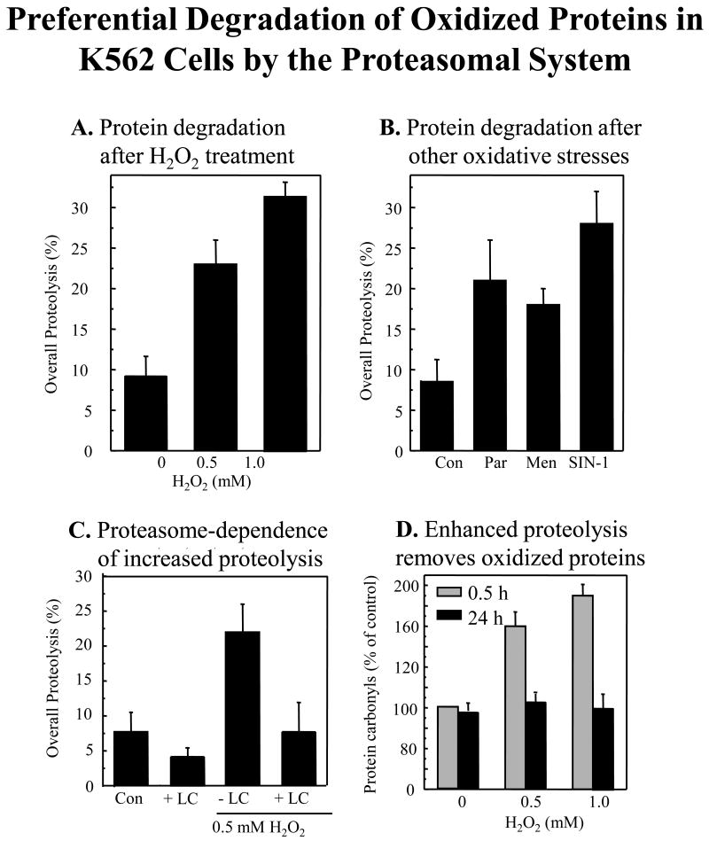
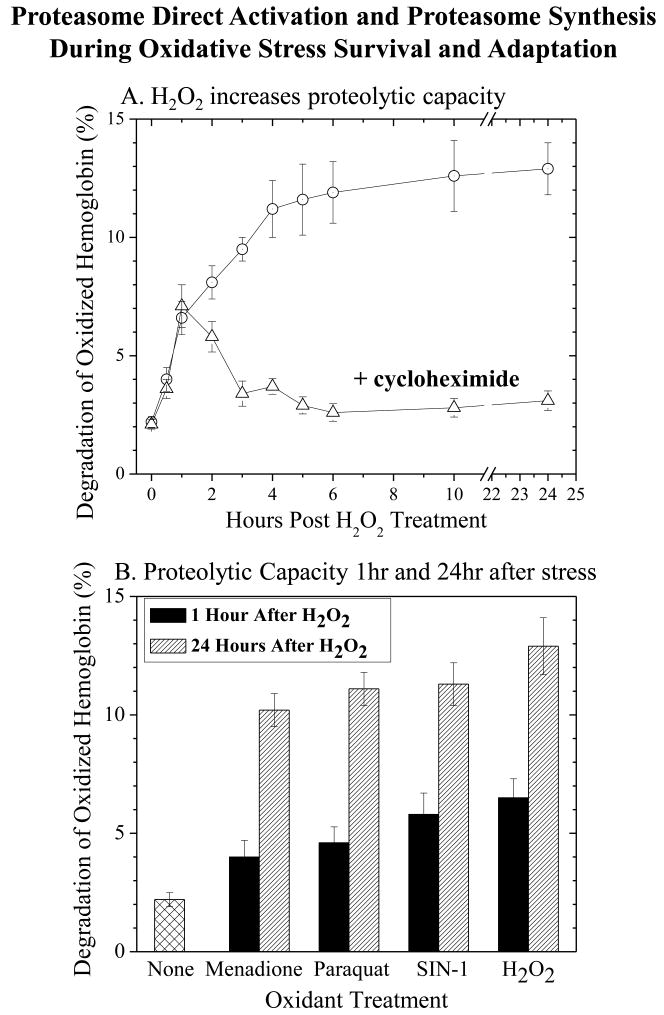
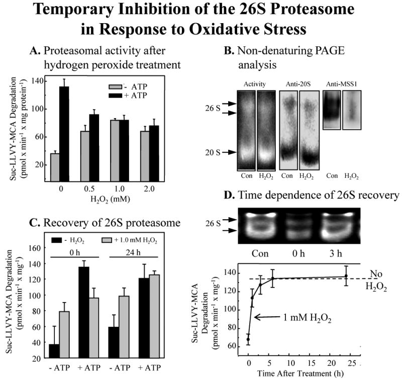
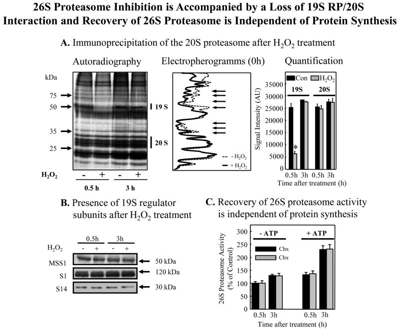
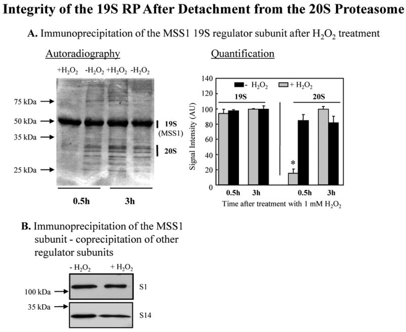
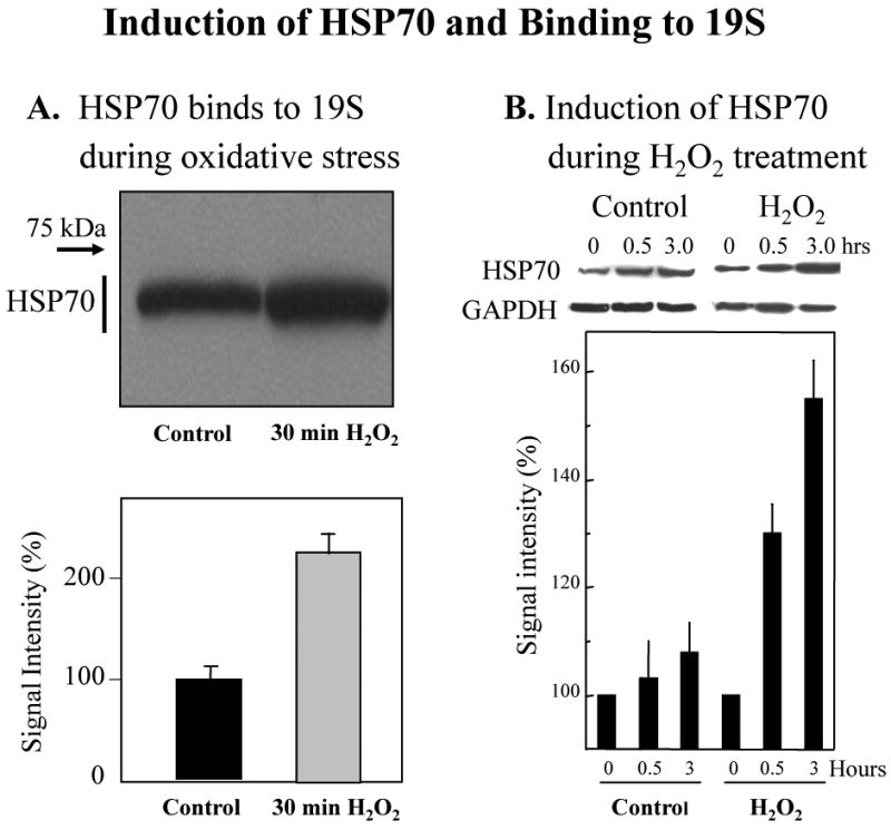
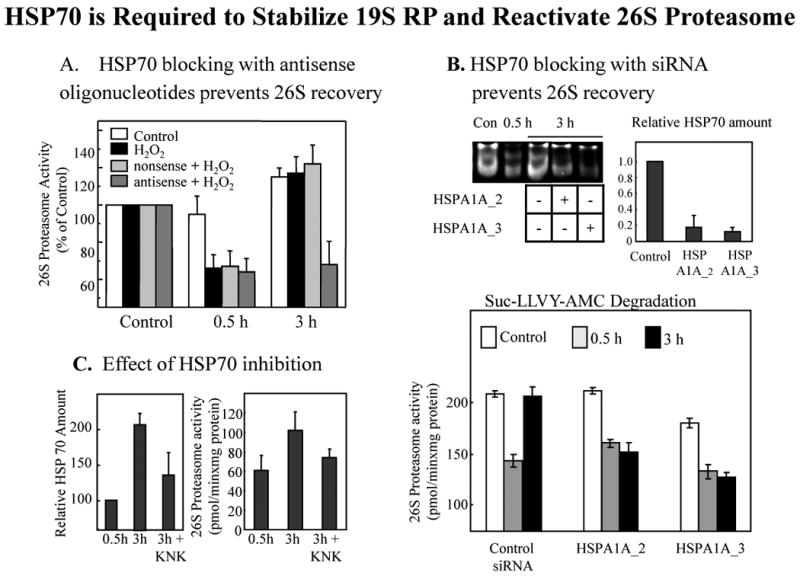
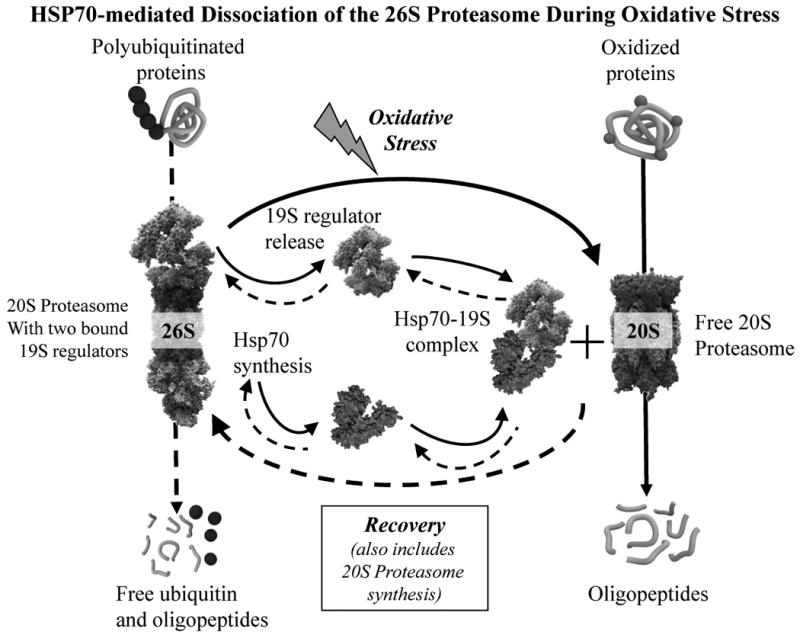
We report an entirely new role for the HSP70 chaperone in dissociating 26S proteasome complexes (into free 20S proteasomes and bound 19S regulators), preserving 19S regulators, and reconstituting 26S proteasomes in the first 1-3h after mild oxidative stress. These responses, coupled with direct 20S proteasome activation by poly(ADP ribose) polymerase in the nucleus and by PA28αβ in the cytoplasm, instantly provide cells with increased capacity to degrade oxidatively damaged proteins and to survive the initial effects of stress exposure. Subsequent adaptive (hormetic) processes (3-24h after stress exposure), mediated by several signal transduction pathways and involving increased transcription/translation of 20S proteasomes, immunoproteasomes, and PA28αβ, abrogate the need for 26S proteasome dissociation. During this adaptive period, HSP70 releases its bound 19S regulators, 26S proteasomes are reconstituted, and ATP-stimulated proteolysis is restored. The 26S proteasome-dependent, and ATP-stimulated, turnover of ubiquitinylated proteins is essential for normal cell metabolism, and its restoration is required for successful stress adaptation.


Figures

Fig. 1
Fig. 1. Preferential degradation of oxidized proteins in K562 cells by the proteasomal system
Panel A. Degradation of metabolically radio-labeled proteins after H2O2 treatment. Endogenous proteins in K562 cells were metabolically radio-labeled with [35S] Met/Cys, in the pulse/chase procedure described in Materials & Methods. Next, 0, 0.5, or 1mM H2O2 was added in PBS, and percent degradation of [35S] cellular proteins was measured after 24 hours. Panel B. Degradation of metabolically radio-labeled proteins after treatment with paraquat, menadione, or SIN-1. Cells were radio-labeled as per Panel A, and percent protein degradation was measured 24 hours after treatment with various oxidant generators; 20 μM paraquat, 20 μM menadione or 1 mM Sin-1, added in PBS. Panel C. Proteasome-dependence of increased proteolysis. The proteasome-dependence of the proteolysis seen in Panels A and B was tested with the proteasome inhibitor, lactacystin. Cells were exposed to 0.5 mM H2O2 as per Panel A, in the presence or absence of lactacystin (LC) at 20μM. Values are means ± SE's, of four experiments with three measurements each. Panel D. Enhanced proteolysis after H2O2 adaptation removes oxidized intracellular proteins. Cells were treated with 0, 0.5, or 1.0 mM H2O2 as per Panel A. Oxidized intracellular proteins were measured by protein carbonyl ELISA (see Materials & Methods) immediately after 0.5 hr of H2O2 treatment, or after 24 hrs. In all panels, oxidant exposures were conducted in 10% of the volume of the original cell suspension and, after 30 min, the remaining 90% of the volume of complete tissue culture medium was re-added. In Panels A, B, and C, all values are means ± SE of four experiments; in Panel D, values are means ± SE of six experiments, each in triplicate.
Fig. 2
Fig. 2. Both direct activiation and de novo synthesis of proteasome occur during adaptation to oxidative stress
Panel A. Cycloheximide effects on increased proteolytic capacity. K562 cells were treated with 0.5 mM H2O2 for 30 min, and cycloheximide (100 g/ml) was then added for incubations lasting from 0.5 hrs to 24 hrs. After various time points over 24 hrs, cell extracts were prepared, and their proteolytic capacity to degrade oxidized [3H] hemoglobin was measured by release of acid-soluble counts (liquid scintillation) as described in Materials & Methods. Panel B. The capacity of cell extracts to degrade oxidized [3H] hemoglobin was measured at both 1 hr, and 24 hrs (both without cycloheximide) after treatment with 0.5 mM hydrogen peroxide, 20 μM paraquat, 20 μM menadione or 1 mM SIN-1, as per Panel A. Values in both panels are means ± SE of three experiments, each in triplicate.
Fig. 3
Fig. 3. Temporary inhibition of the 26S proteasome in response to oxidative stress
Panel A. Loss of ATP-stimulated, 26S proteasome dependent, activity with H2O2 treatment. Human hematopioetic K562 cells were treated with various concentrations of H2O2 for 30 min, as per Fig. 1. Cells were then immediately harvested and lysed, and the extracts were used to measure capacity to degrade the fluorogenic peptide substrate Suc-LLVY-AMC (a measure of proteasomal chymotrypsin-like activity) in the presence or absence of ATP/Mg2+, as described in Materials & Methods. Data are means ± SE, of six experiments, each in triplicate. Panel B. Proteasome activity gel and Western blots. The panel shows representative non-denaturing proteolytic activity gels using Suc-LLVY-AMC as a substrate (left lanes) [13], immunoblots developed with a (Biomol) anti-20S proteasome antibody (middle lanes), and a (Biomol) anti-MSS1 26S proteasomal-subunit-antibody (right lanes). The lysates were harvested 30min after addition of 1.0 mM H2O2. The activity gel background is black, so proteolysis shows as negative staining (light bands) in the region of the two 26S proteasomal bands and the single 20S proteasome band. The activity gel clearly reveals loss of 26S activity, with increase in 20S activity, after 0.5 hr of H2O2 treatment. Panel C. Recovery of ATP-dependent 26S proteasomal activity 24 hours after H2O2 treatment. Suc-LLVY-AMC degradation, in the presence or absence of ATP/Mg2+ was used to measure the recovery of ATP-dependent 26S proteasomal activity, 0 and 24 hours after H2O2 treatment. Panel D. Time dependence of the recovery of 26S protesomal activity. Experiments were performed as per Panel C. The upper part of the panel represents the 26S proteasome activity in an activity stain of a non-denaturing electrophoresis gel. In the lower part of Panel D, the broken line represents 26S proteasomal activity without H2O2 treatment. The solid line connecting data points reports values for cells treated with 1.0 mM hydrogen peroxide. Values in Panels A and C are means ± SE of four experiments, each in triplicate. Values in Panel D are means ± SE of five experiments, each in triplicate.
Fig. 4
Fig. 4. 26S proteasome inhibition is accompanied by a loss of 19SRP/20S interaction and recovery of 26S proteasome is independent of protein synthesis
Panel A. Immunoprecipitation of the 20S proteasome after H2O2 treatment. Human K562 cells were cultured and metabolically radio-labeled in a pulse-chase procedure, with [35S] -Cys/Met (50 μCi/ml or 1 mCi/107 cells) as described in Materials & Methods. Cells were then treated with 1.0 mM H2O2 and harvested at either 0.5 hr or at 3 hrs after treatment. Immunoprecipitation (see Materials & Methods) of 20S proteasome was performed using an anti-20S proteasome-antibody (Biomol, 2 μl antibody per mg cell lysate). The immunoprecipitate was separated on a 12 % PAGE and analyzed by autoradiography. One representative autoradiogram is shown to the left, and its electropherogram is in the middle of the panel; arrows indicate significant differences between control (dotted line) and H2O2 treated (solid line) samples. Quantification of 20S proteasome and 19S regulator bands from six gels (means ± SE) is shown at the right of Panel A. For quantification of 20S proteasome, we used the upper group of 20S proteasome bands, whereas for 19S regulator quantification we used an anti-MSS1 antibody (Biomol) and measured the 51 kDa co-precipitating band of the MSS1 19S regulator particle. Similar results were obtained using 0.5 mM H2O2 treatment (data not shown). Panel B. Preservation of 19 S regulator subunits MSS1, S1, and S14 after H2O2 treatment. Cells were treated with 1.0 mM H2O2 or used as controls, as in Panel A. After 30 min or 3.0 hrs, the levels of 19S regulator subunits was tested by Western blot. No significant changes in MSS1, S1, or S14 intracellular levels were detected at any point, and this analysis was repeated several times with the same results. Panel C. Recovery of 26S proteasome activity is independent of protein synthesis. K562 cells were treated with 1.0 mM H2O2 as per panels A and B. Cycloheximide (100 μg/ml) was then added to half of the cells and cell extracts were prepared. ATP-stimulated 26S proteasome activity was measured in the extracts by degradation of Suc-LLVY-AMC ± ATP/Mg2+ as per Figs. 3C and 3D. Values are means ± SE of four experiments, each in triplicate.
Fig. 5
Fig. 5. Integrity of the 19S regulator particle after dissociation from the 20S proteasome
Human K562 cell were cultivated and proteins were metabolically radio-labeled, in a pulse-chase procedure, with [35S] Met/Cys as described in Fig. 1. Panel A. Immunoprecipitation of the MSS1 19S proteasomal regulator after H2O2 treatment. Cells were incubated for 0.5 or 3.0 hrs after treatment with 1.0 mM H2O2 or used as controls. They were harvested and lysed, and immunoprecipitated (see Materials & Methods) with an anti-MSS1 19S proteasomal subunit regulator antibody (Biomol, at 2 μl/mg cell lysate). The immunoprecipitate was analyzed by PAGE, and bands detected by autoradiography. A representative autoradiogram is shown to the left of the panel, in which the MSS1 band is always strong, but the co-precipitation of several 20S proteasome bands is only seen in control samples, and 3 hr after H2O2 treatment. Quantification of results from six gels (means ± SE) is given to the right of the panel where 20S proteasome co-precipitation with MSS1 is clearly seen to be lost 0.5 hr after H2O2 treatment (P < 0.01), but is recovered by 3 hr. Panel B. Co-precipitation of other 19S proteasome regulator subunits with MSS1. Samples from the experiments in Panel A were immunoprecipitated with the MSS1 antibody (Biomol), and then studied by Western blot with antibodies (biomol) against the S1 and S14 subunits of the 19S proteasome regulator.
Fig. 6
Fig. 6. Binding of HSP70 to the 19S regulator particle and induction of HSP70
Panel A. Binding of HSP70 to the 19S regulator particle. Human K562 cells were used as controls, or were treated for 30 min with 1.0 mM H2O2 as described above. Cells were then lysed, and the extracts were immunoprecipited (see Materials & Methods) with an antibody (Biomol) against the MSS1 subunit of the 19S proteasome regulator particle. Western blots for co-immunoiprecipitation of HSP70 were then performed. HSP70 Western blots utilized a monoclonal anti-HSP70 (human) antibody from Enzo Life Sciences International, Inc. (Plymouth Meeting, PA), product # ADI-SPA-810. According to the manufacturer, this clone92F3A-5 antibody does not cross-react with the constitutive Hsc70 protein; a representative Western blot is shown at the top of Panel A, and quantification of HSP70 bound to 19S regulators is shown at the bottom of the panel as the average of 6 gels (means ± SE). Panel B. Induction of HSP70 during H2O2 treatment. Cells were treated with 1.0 mM H2O2 and collected after 0.5 or 3.0 hrs, or were used as controls. HSP70 protein levels were measured in cell extracts by quantification of Western blots, and data are means ± SE of six experiments. HSP70 Western blots utilized a monoclonal anti-HSP70 (human) antibody from Enzo Life Sciences International, Inc. (Plymouth Meeting, PA), product # ADI-SPA-810. According to the manufacturer, this clone92F3A-5 antibody does not cross-react with the constitutive Hsc70 protein.
Fig. 7
Fig. 7. HSP70 is required to stabilize 19S RP and reactivate 26S proteasome after oxidative stress
Human K562 cells were treated with 1.0mM H2O2, or were used as controls, as above. Panel A. HSP70 antisense oligonucleotides block recovery of 26S proteasome activity after oxidative stress. K562 cells were pre-incubated with either an HSP70 antisense oligonucleotide (5'-CAC CTT GCC GTG CTG GAA-3') or a nonsense (ns) oligonucleotide, in a final concentration of 10 μM as described by Robertson et al. [50] and as previously employed [13, 16]. After 0, 0.5, or 3.0 hrs of H2O2 treatment, cells were harvested and analyzed for proteasomal activity by suc-LLVY-AMC degradation in the presence of ATP; no changes in proteasome activity in the absence of ATP were observed (data not shown). Values are means ± SE of three experiments, each in triplicate. Panel B. HSP70 siRNA blocks recovery of 26S proteasome activity after oxidative stress. Using procedures previously described [1], K562 cells were transfected, using the HiPerFect Transfection Reagent (#301704), with either of two different siRNA's specific for HSP70: Qiagen product # SI00442967, Hs_HSPA1A_2HP (5'-TCC TGT GTT TGC AAT GTT GAA-3') and Qiagen product #SI00442974, Hs_HSPA1A_3HP siRNA (5'-AGA GAT GAA TTT ATA CTG CCA-3') to a final concentration of 10 μM. Qiagen reports that these siRNA's are specific for the inducible HSP70 and will not affect levels of the constitutive HSC70. The actual HSP70 protein knock-down we achieved with these siRNA treatments was measured by Western blot (as per Fig. 6, relative to GAPDH loading controls) and can be seen in the upper right corner of Panel B (values are means ± SE's of six experiments). The upper left corner of Panel B shows a non-denaturing activity gel of suc-LLVY-AMC degradation [13] in the presence of ATP for control cells, cells treated with H2O2 for 0.5 hr, and cells treated with H2O2 for 3 hrs ± the HSPA1A_2 or HSPA1A_3 HSP70 siRNA's. The gel background is black, so proteolysis shows as negative staining (light bands) in the region of the two 26S proteasomal bands, and clearly reveals loss of 26S activity after 0.5 hr of H2O2 followed by recovery at 3 hr, except when HSP70 induction is blocked with HSPA1A_2 or HSPA1A_3 siRNA's. The lower portion of Panel B shows the results of a fluorescence-based suc-LLVY-AMC degradation assay in the presence of ATP, as a more detailed and quantifiable measure of ATP-stimulated 26S proteasomal activity. Proteolysis was measured by fluorescence emission of free AMC in comparison with standards (see Materials & Methods) after 0, 0.5, or 3.0 hrs of H2O2 treatment, in the presence or absence of the HSPA1A_2 or HSPA1A_3 HSP70 siRNA's; values are means ± SE of three experiments, each in triplicate. Panel C. Effect of the HSP70 inhibitor KNK437 on HSP70 levels and proteasome activity. Cells were pretreated with 100 μM KNK437 for 1 h prior to treatment with 1.0 mM hydrogen peroxide. Cell lysates were analyzed 0.5 h and 3 h after H2O2 treatment by immunoblotting for HSP70 amount (as per Fig. 6) in the left side of the panel, and by AMC fluorescence for 26S proteasomal activity in the presence of ATP in the right side of the panel; all values are means ± SE of three experiments, each in triplicate.
Fig. 8
Fig. 8. HSP70-mediated dissociation of the 26S Proteasome during oxidative stress
Scematic representation of the major findings of this study. Oxidative stress causes synthesis of HSP70 which forms complexes with 19S regulator proteins of the 26S proteasome, thus generating extra free 20S proteasomes to degrade oxidized proteins as an initial response to oxidative stress. After about three hours, as cells begin to adapt to the stress, the HSP70-19S regulator complexes dissociate and 26S proteasomes reform. If the stress is relatively mild, and cells can mount an adaptive response (hormesis), de novo 20S proteasome synthesis will also begin, as will synthesis of the immunoproteasome and the Pa28αβ (or 11S) proteasome regulator, all of which can contribute to the continued removal of proteins that were oxidized during the stress [1]. In the Scheme, solid arrows indicate oxidative stress effects whereas dashed arrows represent recovery, or unstressed, processes.

References

    1. Pickering AM, Koop AL, Teoh CY, Ermak G, Grune T, Davies KJA. The immunoproteasome, the 20S proteasome and the PA28alphabeta proteasome regulator are oxidative-stress-adaptive proteolytic complexes. Biochem J. 2010;432:585–594. - PMC - PubMed
    1. Ullrich O, Reinheckel T, Sitte N, Hass R, Grune T, Davies KJA. Poly-ADP ribose polymerase activates nuclear proteasome to degrade oxidatively damaged histones. Proc Natl Acad Sci U S A. 1999;96:6223–6228. - PMC - PubMed
    1. Tanaka K, Kasahara M. The MHC class I ligand-generating system: roles of immunoproteasomes and the interferon-gamma-inducible proteasome activator PA28. Immunol Rev. 1998;163:161–176. - PubMed
    1. Ma CP, Slaughter CA, DeMartino GN. Identification, purification, and characterization of a protein activator (PA28) of the 20 S proteasome (macropain) J Biol Chem. 1992;267:10515–10523. - PubMed
    1. Ahn K, Erlander M, Leturcq D, Peterson PA, Fruh K, Yang Y. In vivo characterization of the proteasome regulator PA28. J Biol Chem. 1996;271:18237–18242. - PubMed

Publication types

MeSH terms

Substances

LinkOut - more resources