Review
. 2011 Nov-Dec;33(6):631-40.
doi: 10.1016/j.ntt.2011.06.006. Epub 2011 Jul 13.

The negative side of retinoic acid receptors

Affiliations
Review

The negative side of retinoic acid receptors

Elwood Linney et al. Neurotoxicol Teratol. 2011 Nov-Dec.

Abstract

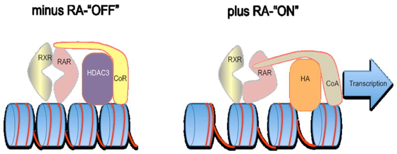
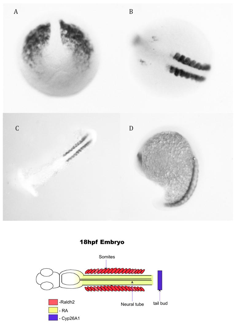
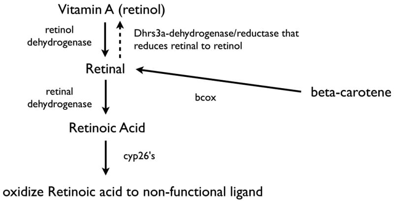
This is a review of research that supports a hypothesis regarding early restriction of gene expression in the vertebrate embryo. We hypothesize that vertebrate retinoic acid receptors (RARs for several vertebrates but rars for zebrafish) are part of an embryonic, epigenetic switch whose default position, at the time of fertilization is "OFF". This is due to the assemblage of a rar-corepressor-histone deacetylase complex on retinoic acid response elements (RAREs) in regulatory regions of a subset of genes. In addition, selective and precise allocation of retinoic acid during early development through the interaction of Phase I enzymes throws the switch "ON" in a predictable, developmental manner. We are proposing that this is a basic, early embryonic switch that can cause the initiation of cascades of gene expression that are responsible for at least some early, diversification of cell phenotypes. Dehydrogenases and a subset of cytochrome p450 genes (cyp26a1, cyp26b1, and cyp26c1) play the major role in providing the retinoic acid and limiting its access. We also suggest that this mechanism may be playing a significant role in the repression of genes in undifferentiated stem cells.


Conflict of interest statement

Conflict of Interest: A conflicting interest exists when professional judgement concerning a primary interest (such as patient's welfare or the validity of research) may be influenced by a secondary interest (such as financial gain or personal rivalry). It may arise for the authors when they have financial interest that may influence their interpretation of their results or those of others. Examples of potential conflicts of interest include employment, consultancies, stock ownership, honoraria, paid expert testimony, patent applications/registrations, and grants or other funding.

Figures

Figure 1
Figure 1
A model of the epigenetic switch. To the left is the “OFF” position where the RAR/RXR heterodimer is binding to a Retinoic Acid Response Element (RARE) in the absence of the retinoic acid ligand. This creates a corepressor binding site on the RAR (CoR binding site) and the corepressor has a histone deacetylase (HDAC3) binding site. As described in the text, this causes both a condensation of the chromatin and a repression of transcription. To the right is the “ON” position. When the retinoic acid ligand (RA) binds to the retinoic acid receptor (RAR) the receptor goes through an allosteric change eliminating the corepressor binding site and creating an association site for a coactivator (CoA). This also brings in a histone acetyltransferase (HA) that helps to open up the chromatin. The coactivator provides the association with the transcription complex to facilitate transcription. This model is derivative of several studies on individual components of the RAR corepressor complex in cell culture.
Figure 2
Figure 2
Retinoic acid receptors, domains, and response elements. A. The basic domain structure of the members of the steroid superfamily including the retinoic acid receptors (RARs) and the retinoid-X receptors (RXRs). B. A comparison of the identified mouse/human RARs and RXRs and the zebrafish rars and rxrs. Because the fish went through an additional genomic duplication, some genes in zebrafish have duplicates, such as the rar a and rar g genes. In addition no rar b genes have been identified for zebrafish. C. The DNA binding sequences that RAR/RXR heterodimers bind to are named RAREs. In the genome, these can take many forms but in many cases they are direct repeats. A prototypical RARE is shown and based upon a study by (Umesono K, et al. 1991).
Figure 3
Figure 3
Identification of mRNAs for z smrt, z ncor, z rar aa and z rar ab in 8-cell embryos before the embryonic genome turns on. This figure plus separate in situ hybridizations with z rxr ab, z rxr ga, zebrafish histone deacetylase 3 (z hdac3) in 8-cell embryos and oocytes support the hypothesis that the molecular machinery for repression is available at the time of fertilization. The z rar aa and z rar ab in situs are consistent with PCR data of Waxman and Yelon (Waxman JS and Yelon D 2007).
Figure 4
Figure 4
Transgenic indicator embryos for the developmental detection of retinoic acid receptor activity. The transgenes are identical except for the reporter. Both have 3 RARE elements coupled the Herpes simplex virus thymidine kinase promoter. The mouse transgene (left) is driving the bacterial beta-galactosidase gene while the zebraifish transgene is driving a Green Fluorescent Protein gene. Note that both are expressed in the neural tube. For details of developmental expression see (Balkan W, et al. 1992) and (Perz-Edwards A, et al. 2001) studies.
Figure 5
Figure 5
Raldh2 in situ hybridization at different developmental stages. A. Tailbud stage. B.10 somite stage-note the distinct localization in the somites adjacent to the neural tube. C.14 somite stage, flatmount. D. 20 somites stage, side view. The cartoon below represents the expression of the raldh2 in the somites—its expression in the brain is not included in the cartoon.
Figure 6
Figure 6
Retinoid pathways for the synthesis and oxidation of retinoic acid. Retinoic acid comes from either vitamin A or beta-carotene that is converted to retinal via retinol dehydrogenases. Retinoic acid is produced by the action of one of several retinal dehydrogenases, the major early embryonic enzyme being retinaldehyde dehydrogenase 2.
Figure 7
Figure 7
Cyp26b1 (blue)/Raldh2 (red) expression at 30h and 48hpf. Lateral views of in situ hybridization localization of the cyp26b1 and ralhd2 mRNAs in A. 30hpf embryo and B. 48hpf embryo. Note the discrete localization of cyp26b1 in the brain and the expression of raldh2 in the eye. More detailed expression of both of these can be found in the (Dobbs-McAuliffe B, et al. 2004) and (Zhao Q, et al. 2005) studies from this laboratory and a more detailed study showed the expression of all three of the cyp26 mRNAs can be found in (Hernandez RE, et al. 2007) study.
Figure 8
Figure 8
A retinoic acid pulse increases the number of neurons in the embryo. Upper right is the experimental timing. 10-6 M retinoic acid was used to exposed embryos from 10hpf to 14hpf. They were washed out of the retinoic acid, allowed to grow to 24hpf fertilization, fixed and quantified for neuron numbers from somites 8-17.

References

    1. Alexa K, Choe SK, Hirsch N, Etheridge L, Laver E, Sagerstrom CG. Maternal and zygotic aldh1a2 activity is required for pancreas development in zebrafish. PLoS One. 2009;4:e8261. - PMC - PubMed
    1. Balciunas D, Wangensteen KJ, Wilber A, Bell J, Geurts A, Sivasubbu S, Wang X, Hackett PB, Largaespada DA, McIvor RS, et al. Harnessing a high cargo-capacity transposon for genetic applications in vertebrates. PLoS Genet. 2006;2:e169. - PMC - PubMed
    1. Balkan W, Colbert M, Bock C, Linney E. Transgenic indicator mice for studying activated retinoic acid receptors during development. Proc Natl Acad Sci U S A. 1992;89:3347–51. - PMC - PubMed
    1. Bertrand S, Thisse B, Tavares R, Sachs L, Chaumot A, Bardet PL, Escriva H, Duffraisse M, Marchand O, Safi R, et al. Unexpected novel relational links uncovered by extensive developmental profiling of nuclear receptor expression. PLoS Genet. 2007;3:e188. - PMC - PubMed
    1. Castillo HA, Cravo RM, Azambuja AP, Simoes-Costa MS, Sura-Trueba S, Gonzalez J, Slonimsky E, Almeida K, Abreu JG, de Almeida MA, et al. Insights into the organization of dorsal spinal cord pathways from an evolutionarily conserved raldh2 intronic enhancer. Development. 2010;137:507–18. - PMC - PubMed

Publication types

MeSH terms