. 2011 Jul 21;10(1):65-74.
doi: 10.1016/j.chom.2011.06.006.

The membrane-bound transcription factor CREB3L1 is activated in response to virus infection to inhibit proliferation of virus-infected cells

Affiliations

The membrane-bound transcription factor CREB3L1 is activated in response to virus infection to inhibit proliferation of virus-infected cells

Bray Denard et al. Cell Host Microbe. .

Abstract

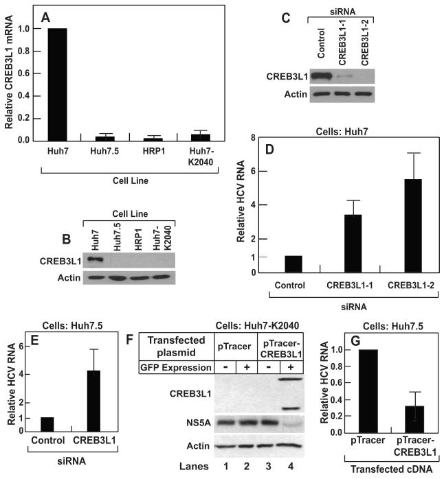
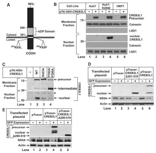
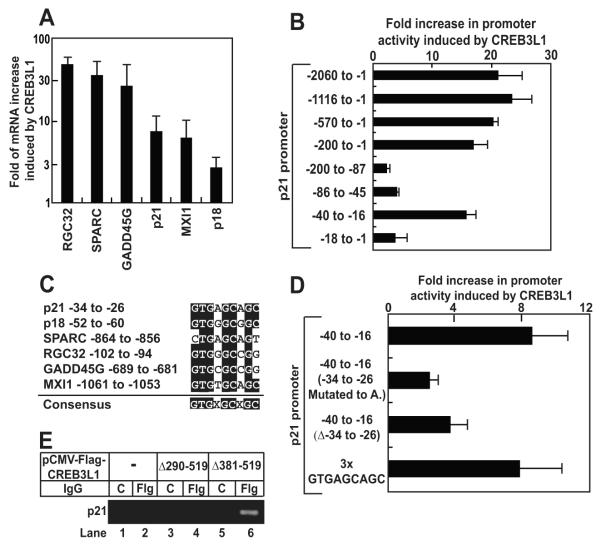
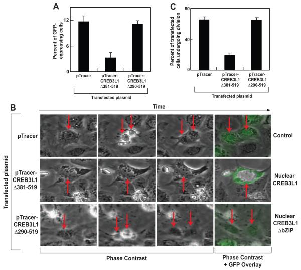
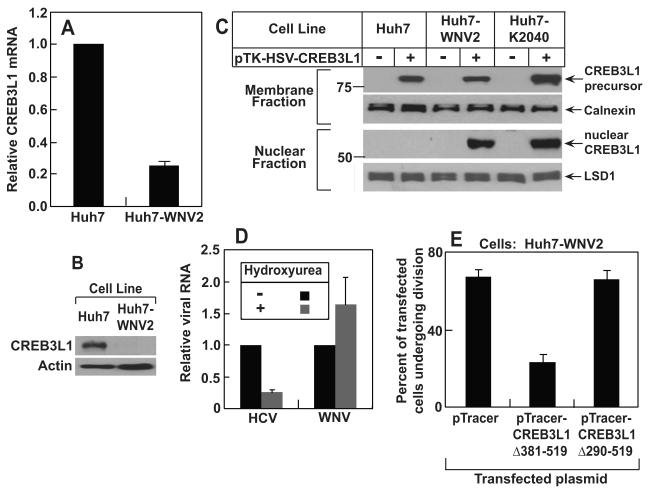
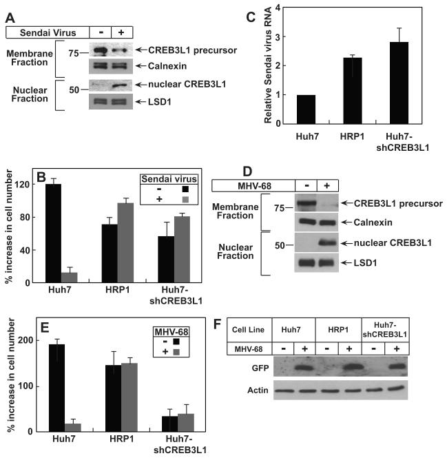
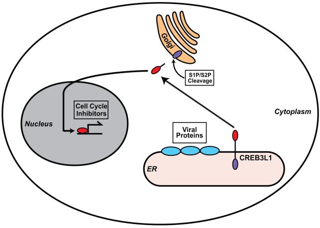
CREB3L1/OASIS is a cellular transcription factor synthesized as a membrane-bound precursor and activated by regulated intramembrane proteolysis in response to stimuli like ER stress. Comparing gene expression between Huh7 subclones that are permissive for hepatitis C virus (HCV) replication versus the nonpermissive parental Huh7 cells, we identified CREB3L1 as a host factor that inhibits proliferation of virus-infected cells. Upon infection with diverse DNA and RNA viruses, including murine γ-herpesvirus 68, HCV, West Nile virus (WNV), and Sendai virus, CREB3L1 was proteolytically cleaved, allowing its NH(2) terminus to enter the nucleus and induce multiple genes encoding inhibitors of the cell cycle to block cell proliferation. Consistent with this, we observed a necessity for CREB3L1 expression to be silenced in proliferating cells that harbor replicons of HCV or WNV. Our results indicate that CREB3L1 may play an important role in limiting virus spread by inhibiting proliferation of virus-infected cells.


Figures

Figure 1
Figure 1. CREB3L1 inhibits HCV replication
(A) RT-QPCR quantification of CREB3L1 mRNA in indicated cells with the value in Huh7 cells set to 1. (B) Immunoblot analysis of CREB3L1 protein in indicated cells. (C and D) On day 0, Huh7 cells were seeded at 1×10⁵/60 mm dish. On day 1, they were transfected with indicated siRNA (20 pmol/dish). On day 3, these cells were transfected with the HCV replicon RNA (HP, 0.5 μg/dish). On day 4, the cells were harvested and the amount of CREB3L1 protein was determined by immunoblot analysis (C). HCV RNA was quantified by RT-QPCR, with the amount of HCV RNA in cells treated with the control siRNA set to 1 (D). (E) Huh7.5 cells were seeded and transfected with indicated siRNA as described in (C). On day 3, the cells were infected with the JFH strain of HCV virion. On day 4, the cells were harvested and the amount of HCV RNA was quantified as described in (C). (F) On day 0, Huh7-K2040 cells were seeded at 7×10⁵/60mm dish. On day 1, they were transfected with 0.5 μg of a plasmid derived from pTracer as indicated. On day 2, the cells were trypsinized and sorted through FACS based on expression of GFP. Lysates of the cells with or without GFP expression were subject to immunoblot analysis with indicated antibodies. (G) Huh7.5 cells transfected with the indicated plasmid were isolated from untransfected Huh7.5 cells based on GFP expression as described in (D), and seeded at 2×10⁵/60mm dish. They were infected by the JFH strain of HCV virion 24 h later. Following incubation for 3 days, HCV RNA in the cells was quantified by RT-QPCR, with the value in cells transfected with the empty pTracer set at 1. (A-G), Bar graphs are reported as mean ± S.D. (n=3).
Figure 2
Figure 2. CREB3L1 undergoes RIP in HCV-infected cells
(A) Schematic diagram of CREB3L1. (B and C) On day 0, indicated cells (B) or Huh7-K2040 cells (C) were seeded at 4×10⁵/60 mm dish. On day 1, they were transfected with 50 ng of indicated plasmids. On day 2, the cells were harvested and separated into nuclear and membrane fractions followed by immunoblot analysis with anti-HSV. Asterisk (*) denotes a cross-reactive band. Immunoblots of calnexin and Lysine-specific demethylase 1 (LSD1) served as loading controls for membrane and nuclear fractions, respectively. (D and E) Huh7-K2040 cells transfected with the indicated plasmid were analyzed as described in Figure 1F.
Figure 3
Figure 3. Nuclear form of CREB3L1 activates genes that inhibit the cell cycle
(A) Huh7-K2040 cells were seeded, transfected with pTracer or pTracer-CREB3L1(Δ381-519) as described in Figure 1D. The transfected cells were isolated based on GFP expression as described in Figure 1D. The amount of indicated mRNA was quantified by RT-QPCR. Fold induction of indicated mRNA by CREB3L1(Δ381-519) was determined by setting the amount of the mRNA in cells transfected with the control plasmid pTracer at 1. Result are reported as mean ± S.D. (n=3). (B and D) On day 0, Huh7-K2040 cells were seed at 7×10⁵/60 mm dish. On day 1, the cells were transfected with a firefly luciferase reporter plasmid containing the indicated region of p21 promoter (1 μg/dish) and a plasmid encoding renilla luciferase driven by the constitutive CMV promoter (0.5 μg/dish) in the absence or presence of cotransfection of pCMV-CREB3L1(Δ381-519) (0.5 μg/dish). On day 2, luciferase activity was measured and the promoter activity was determined by firefly luciferase activity normalized against renilla luciferase activity to control for transfection efficiency. Fold increase in the promoter activity in cells transfected with CREB3L1(Δ381-519) compared to those mock-transfected was presented. Result are reported as mean ± S.D. (n=3). (C) Alignment of −34 to −26 of the p21 promoter region with promoter sequences from indicated genes. (E) On day 0, Huh7-K2040 cells were seeded at 2.5×10⁵/60 mm dish. On day 3, they were transfected with 0.5 μg of indicated plasmids. On day 4, the cells were harvested and the cell lysate was immunoprecipitated with control IgG (C) or anti-Flag IgG (Flg) followed by ChIP analysis as described in Experimental Procedures.
Figure 4
Figure 4. Nuclear form of CREB3L1 inhibits cell proliferation
(A) Huh7-K2040 cells were seeded and transfected with the indicated plasmid as described in Figure 1D. Three days after the transfection, percentage of the cells expressing GFP encoded by the transfected plasmid was determined through FACS analysis. Results are reported as mean ± S.D. (n=3). (B) On day 0, Huh7-K2040 cells were seeded at 2.5×10⁵ per well of a 6-well plate. On day 1, they were transfected with 0.25 μg of the indicated plasmid. On day 2, the cells were subjected to time-lapse imaging analysis as described in Experimental Procedures. On day 4 (48 h later), GFP fluorescence images were taken. Representative time-lapse images of these cells were shown, with arrows indicating transfected cells and their daughter cells determined by their expression of GFP at the end of the imaging at 48 h. (C) Quantification of transfected cells that went through cell divisions in (B). On average, 30 cells transfected with each plasmid were counted in each experiment. Results are reported as mean ± S.D. (n=3).
Figure 5
Figure 5. RIP of CREB3L1 inhibits proliferation of cells infected by WNV
(A) RT-QPCR quantification of CREB3L1 mRNA in indicated cells with the value in Huh7 cells set to 1. Results are reported as mean ± S.D. (n=3). (B) Immunoblot analysis of CREB3L1 protein precursor in indicated cells (C) Proteolytic cleavage of CREB3L1 was analyzed as described in Figure 2B. (D) On day 0, Huh7-K2040 and Huh7-WNV2 cells were seeded at 2×10⁵/60 mm dish. On day 1, the cells were treated with or without 10 μM hydroxyurea. On day 5, cells were harvested for quantification of HCV and WNV RNA in Huh7-K2040 and Huh7-WNV2 cells, respectively, by RT –QPCR, with the values in cells that were not treated with the drug set to 1. Results are reported as mean ± S.D. (n=3). (E) Quantification of Huh7-WNV2 cells transfected with indicated plasmid that went through cell divisions was carried out as described in Figure 4C.
Figure 6
Figure 6. CREB3L1 prevents proliferation of cells infected by Sendai Virus and MHV-68
(A) On day 0, Huh7 cells were seeded at 4×10⁵/60 mm dish. On day 1, the cells were infected with Sendai virus. On day 2, the cells were fractionated into nuclear and membrane fractions and the cleavage of CREB3L1 was determined by immunoblot analysis with anti-CREB3L1. (B and C) On day 0, Huh7, HRP-1 and Huh7-shCREB3L1 cells were seeded at 2×10⁵/60 mm dish. On day 1, triplicate dishes of the cells were harvested for cell counting. The remaining cells were infected with Sendai virus as indicated. On day 2 (24 h later), triplicate dishes of these cells were harvested and the number of cells in each plate was quantified. The percentage of indicated cells in each plate increased on day 2 compared to that on day 1 is presented (B). Sendai virus RNA in the cells infected by the virus was quantified by RT-QPCR with the value in Huh7 cells set to 1 (C). (D) Proteolytic cleavage of CREB3L1 was analyzed as in (A) except that the cells were infected by MHV-68 instead of Sendai virus. (E and F) Indicated cells were seeded and treated as described in (B) except that the cells were infected by MHV-68 instead of Sendai virus. Proliferation of the cells was determined as described in (B). Cellular lysate was subjected to immunoblot analysis with anti-GFP (F). (A-F) Bar graphs are reported as mean ± S.D. (n=3).
Figure 7
Figure 7. A model illustrating the role of CREB3L1 in limiting HCV infection
CREB3L1 is synthesized as a membrane-bound precursor. In cells infected by virus, CREB3L1 is activated through RIP, presumably through ER stress caused by expression of ER-associated viral proteins. The proteolytic cleavage allows the NH2-terminal domain of the protein to enter the nucleus, where it activates transcription of genes encoding cell cycle inhibitors to block proliferation of the cells infected by the virus.

References

    1. Adams CM, Reitz J, De Brabander JK, Feramisco JD, Li L, Brown MS, Goldstein JL. Cholesterol and 25-hydroxycholesterol inhibit activation of SREBPs by different mechanisms, both involving SCAP and Insigs. J. Biol. Chem. 2004;279:52772–52780. - PubMed
    1. Appel N, Schaller T, Penin F, Bartenschlager R. From structure to function: new insights into hepatitis C virus RNA replication. J. Biol. Chem. 2006;281:9833–9836. - PubMed
    1. Binder M, Kochs G, Bartenschlager R, Lohmann V. Hepatitis C virus escape from the interferon regulatory factor 3 pathway by a passive and active evasion strategy. Hepatology. 2007;46:1365–1374. - PubMed
    1. Blight KJ, McKeating JA, Rice CM. Highly permissive cell lines for subgenomic and genomic hepatitis C virus RNA replication. J. Virol. 2002;76:13001–13014. - PMC - PubMed
    1. Bradshaw AD, Sage EH. SPARC, a matricellular protein that functions in cellular differentiation and tissue response to injury. J Clin Invest. 2001;107:1049–1054. - PMC - PubMed

Publication types

MeSH terms

Substances

Associated data