. 2011 Sep 16;286(37):32672-83.
doi: 10.1074/jbc.M111.236265. Epub 2011 Jul 15.

The F-BAR protein Rapostlin regulates dendritic spine formation in hippocampal neurons

Affiliations

The F-BAR protein Rapostlin regulates dendritic spine formation in hippocampal neurons

Yohei Wakita et al. J Biol Chem. .

Abstract

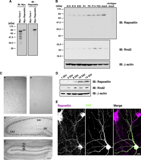
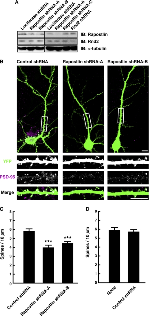
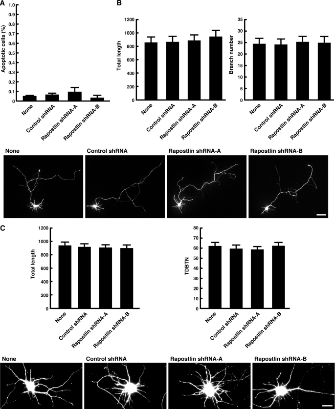
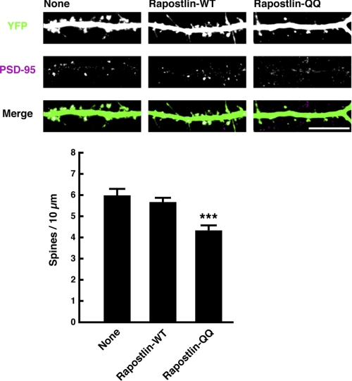
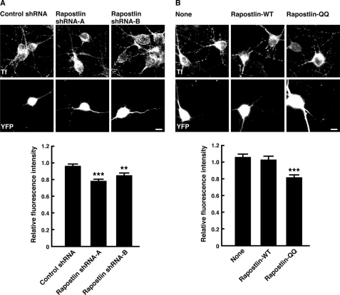
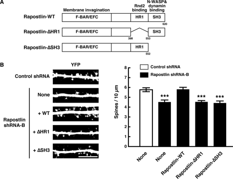
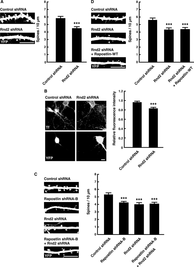
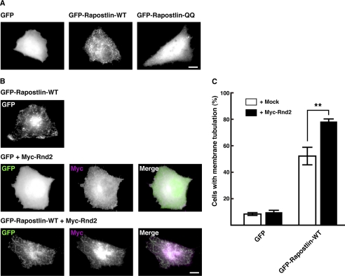
Pombe Cdc15 homology proteins, characterized by Fer/CIP4 homology Bin-Amphiphysin-Rvs/extended Fer/CIP4 homology (F-BAR/EFC) domains with membrane invaginating property, play critical roles in a variety of membrane reorganization processes. Among them, Rapostlin/formin-binding protein 17 (FBP17) has attracted increasing attention as a critical coordinator of endocytosis. Here we found that Rapostlin was expressed in the developing rat brain, including the hippocampus, in late developmental stages when accelerated dendritic spine formation and maturation occur. In primary cultured rat hippocampal neurons, knockdown of Rapostlin by shRNA or overexpression of Rapostlin-QQ, an F-BAR domain mutant of Rapostlin that has no ability to induce membrane invagination, led to a significant decrease in spine density. Expression of shRNA-resistant wild-type Rapostlin effectively restored spine density in Rapostlin knockdown neurons, whereas expression of Rapostlin deletion mutants lacking the protein kinase C-related kinase homology region 1 (HR1) or Src homology 3 (SH3) domain did not. In addition, knockdown of Rapostlin or overexpression of Rapostlin-QQ reduced the uptake of transferrin in hippocampal neurons. Knockdown of Rnd2, which binds to the HR1 domain of Rapostlin, also reduced spine density and the transferrin uptake. These results suggest that Rapostlin and Rnd2 cooperatively regulate spine density. Indeed, Rnd2 enhanced the Rapostlin-induced tubular membrane invagination. We conclude that the F-BAR protein Rapostlin, whose activity is regulated by Rnd2, plays a key role in spine formation through the regulation of membrane dynamics.


Figures

FIGURE 1.
FIGURE 1.
Expression and distribution of Rapostlin in rat brain. A, specificity of the antibody against Rapostlin. Cell lysates from HEK293T cells transfected with Myc-tagged Toca-1 or Rapostlin were immunoblotted (IB) with an antibody against Myc or Rapostlin. B, immunoblot analysis of Rapostlin and Rnd2 from rat brain lysates at various ages. Lysates (50 μg of total protein) of rat brain at the indicated ages were subjected to immunoblotting with the antibody against Rapostlin or Rnd2. We also used the antibody against Rapostlin preincubated with excess antigenic peptide to confirm the specificity of the immunoreactivity observed. The level of β-actin was also analyzed as a loading control. C, immunohistochemistry was performed with the antibody against Rapostlin for coronally sectioned P14 rat cerebral cortex (a and b), hippocampus (c), and cerebellum (d). No immunoreactivity was observed in the section adjacent to a when the antibody was preincubated with an excess amount of antigenic peptide (b). DG, dentate gyrus; GL, granular layer; PL, Purkinje layer; ML, molecular layer. Scale bar = 200 μm. D, expression of Rapostlin and Rnd2 in developing rat hippocampal cell cultures. Lysates of hippocampal cell cultures at various stages were subjected to immunoblotting with the antibody against Rapostlin, Rnd2, or β-actin. E, staining with Rapostlin antibody in primary cultured rat hippocampal neurons at 15 DIV. YFP transfection was used to visualize cell morphology. Scale bar = 10 μm.
FIGURE 2.
FIGURE 2.
Knockdown of Rapostlin decreases spine density in primary cultured rat hippocampal neurons. A, cell lysates from neurons transfected with the luciferase shRNA, Rapostlin shRNA, or Rnd2 shRNA expression vector were subjected to immunoblotting (IB) with the antibody against Rapostlin, Rnd2, or α-tubulin to detect endogenous proteins. B, neurons were transiently transfected with control shRNA (Rapostlin shRNA-C), Rapostlin shRNA-A, or Rapostlin shRNA-B and YFP at 11 DIV, and then they were fixed at 15 DIV and stained with the antibody against PSD-95, a postsynaptic marker protein. The transfected cells are shown by the fluorescence of YFP. The three lower panels show enlargements of the boxed regions in the top panel. The top and bottom panels show the merge of the two images with YFP (green) and PSD-95 (magenta). Scale bars = 10 μm. C and D, quantification of the effect of Rapostlin knockdown on spine density in neurons. The number of spines within dendritic segments of 100–200 μm in YFP-positive neurons was counted, and the spine density was calculated (spines/10 μm dendritic shaft). At least 15 neurons were collected per construct from three experiments, and data represent mean ± S.E. ***, p < 0.01 (versus control shRNA, n = 45, t test).
FIGURE 3.
FIGURE 3.
Knockdown of Rapostlin had no effects on axon and dendrite morphology. A, knockdown of Rapostlin does not induce apoptosis. Neurons were transiently transfected with shRNA and YFP at 11 DIV, fixed at 15 DIV, and then TUNEL assays were performed. TUNEL and YFP double-positive cells were scored for TUNEL-positive nuclei, and data represent mean ± S.E. from three independent experiments. B, quantification of axon morphology. Neurons were transiently transfected with shRNA and YFP at 2 DIV, and then they were fixed at 4 DIV. The axons were identified by staining with the axonal marker Tau-1 (data not shown). Scale bar = 50 μm. The axon length and branch number of transfected neurons were measured. Processes longer than 5 μm were considered branches. At least 15 neurons were collected per construct from three experiments, and data represent mean ± S.E. C, quantification of dendrite morphology. Neurons were transiently transfected with shRNA and YFP at 5 DIV, and then they were fixed at 7 DIV. The dendrites were identified by staining with the dendritic marker MAP2 (data not shown). The total length of dendrites and total dendritic branch tip number (TDBTN) of transfected neurons were measured. Dendritic tips were scored when they were longer than 5 μm. At least 15 neurons were collected per construct from three experiments, and data represent mean ± S.E. Scale bar = 20 μm.
FIGURE 4.
FIGURE 4.
Overexpression of Rapostlin-QQ decreases spine density in primary cultured rat hippocampal neurons. Neurons were transiently transfected with the indicated plasmids at 11 DIV, and then they were fixed at 15 DIV and stained with the antibody against PSD-95. Scale bar = 10 μm. For quantification, the number of spines within dendritic segments of 100–200 μm in YFP-positive neurons was counted, and the spine density was calculated (spines/10 μm dendritic shaft). At least 15 neurons were collected per construct from three experiments, and data represent mean ± S.E. ***, p < 0.01 (versus none, n = 45, t test).
FIGURE 5.
FIGURE 5.
Knockdown of Rapostlin and overexpression of Rapostlin-QQ suppress the uptake of transferrin in primary cultured rat hippocampal neurons. A, neurons were transiently transfected with shRNA and YFP at 7 DIV. After 3 days, they were incubated with Alexa Fluor 594-transferrin for 10 min at 37 °C. The images of Alexa Fluor 594-transferrin (Tf, upper panels) and YFP (lower panels) are shown. Relative fluorescence intensity was determined from the average fluorescence intensity of Alexa Fluor 594-transferrin in dendrites and the soma of YFP-positive neurons normalized to that of YFP-negative untransfected neurons in the same field. At least 20 neurons were collected per construct from three experiments, and data represent mean ± S.E. **, p < 0.01; ***, p < 0.001 (versus control shRNA, n = 60, t test). B, neurons were transiently transfected with Rapostlin-WT or Rapostlin-QQ and YFP at 7 DIV. After 3 days, they were incubated with Alexa Fluor 594-transferrin for 10 min at 37 °C. ***, p < 0.001 (versus none, n = 45, t test). Scale bars = 10 μm.
FIGURE 6.
FIGURE 6.
The HR1 and SH3 domains are involved in the Rapostlin-mediated spine formation. A, the domain structure of Rapostlin. B, primary cultured rat hippocampal neurons were transiently cotransfected with the indicated plasmids at 11 DIV and fixed at 15 DIV. Scale bar = 10 μm. For quantification, the number of spines within dendritic segments of 100–200 μm in YFP-positive neurons was counted, and the spine density was calculated (spines/10 μm dendritic shaft). At least 15 neurons were collected per construct from three experiments, and data represent mean ± S.E. ***, p < 0.001 (versus control shRNA, n = 45, t test).
FIGURE 7.
FIGURE 7.
Knockdown of Rnd2 suppresses spine formation and transferrin uptake in primary cultured rat hippocampal neurons. A, neurons were transiently transfected with control or Rnd2 shRNA and YFP at 11 DIV, and then they were fixed at 15 DIV and stained with the antibody against PSD-95. Scale bar = 10 μm. For quantification, the number of spines within dendritic segments of 100–200 μm in YFP-positive neurons was counted, and the spine density was calculated (spines/10 μm dendritic shaft). At least 15 neurons were collected per construct from three experiments, and data represent mean ± S.E. ***, p < 0.001 (versus control shRNA, n = 45, t test). B, neurons were transiently transfected with control or Rnd2 shRNA and YFP at 7 DIV. After 3 days, they were incubated with Alexa Fluor 594-transferrin for 10 min at 37 °C. The images of Alexa Fluor 594-transferrin (Tf, upper panels) and YFP (lower panels) are shown. Scale bar = 10 μm. Relative fluorescence intensity was determined from the average fluorescence intensity of Alexa Fluor 594-transferrin in dendrites and the soma of YFP-positive neurons normalized to that of YFP-negative untransfected neurons in the same field. At least 20 neurons were collected per construct from three experiments, and data represent mean ± S.E. ***, p < 0.001 (versus control shRNA, n = 60, t test). C and D, neurons were transiently transfected with the indicated plasmids at 11 DIV, and then they were fixed at 15 DIV. Scale bar = 10 μm. For quantification, the number of spines within dendritic segments of 100–200 μm in YFP-positive neurons was counted, and the spine density was calculated (spines/10 μm dendritic shaft). At least 15 neurons were collected per construct from three experiments, and data represent mean ± S.E. ***, p < 0.001 (versus control shRNA, n = 45, t test).
FIGURE 8.
FIGURE 8.
Effect of coexpression of Rnd2 on Rapostlin-induced plasma membrane tubulation in HeLa cells. A, HeLa cells were transfected with GFP, GFP-Rapostlin, or GFP-Rapostlin-QQ, and the images of GFP are shown. B, HeLa cells were cotransfected with GFP or GFP-tagged Rapostlin with Myc-tagged Rnd2 and stained with the antibody against Myc. The right panels show the merge of the two images with GFP (green) and Myc (magenta). Scale bars = 20 μm. C, quantification of the effect of Rnd2 expression on Rapostlin-induced membrane tubulation in HeLa cells. Cells with membrane tubulation were quantified, and results were scored as a percentage of the number of the transfected cells. Results are the means ± S.E. of at least three independent experiments in which more than 50 cells were counted. **, p < 0.01 (versus + mock, t test).

Similar articles

Cited by

References

    1. Hall A. (1998) Science 279, 509–514 - PubMed
    1. Luo L. (2000) Nat. Rev. Neurosci. 1, 173–180 - PubMed
    1. Negishi M., Katoh H. (2002) J. Biochem. 132, 157–166 - PubMed
    1. Burridge K., Wennerberg K. (2004) Cell 116, 167–179 - PubMed
    1. Aspenström P., Fransson A., Richnau N. (2006) Trends Biochem. Sci. 31, 670–679 - PubMed

Publication types

MeSH terms

LinkOut - more resources