. 2011 Sep 9;286(36):31598-609.
doi: 10.1074/jbc.M110.199430. Epub 2011 Jul 15.

ZF21 protein, a regulator of the disassembly of focal adhesions and cancer metastasis, contains a novel noncanonical pleckstrin homology domain

Affiliations

ZF21 protein, a regulator of the disassembly of focal adhesions and cancer metastasis, contains a novel noncanonical pleckstrin homology domain

Makoto Nagano et al. J Biol Chem. .

Abstract

Directional migration of adherent cells on an extracellular matrix requires repeated formation and disassembly of focal adhesions (FAs). Directional migration of adherent cells We have identified ZF21 as a regulator of disassembly of FAs and cell migration, and increased expression of the gene has been linked to metastatic colon cancer. ZF21 is a member of a protein family characterized by the presence of the FYVE domain, which is conserved among Fab1p, YOPB, Vps27p, and EEA1 proteins, and has been shown to mediate the binding of such proteins to phosphoinositides in the lipid layers of cell membranes. ZF21 binds multiple factors that promote disassembly of FAs such as FAK, β-tubulin, m-calpain, and SHP-2. ZF21 does not contain any other known protein motifs other than the FYVE domain, but a region of the protein C-terminal to the FYVE domain is sufficient to mediate binding to β-tubulin. In this study, we demonstrate that the C-terminal region is important for the ability of ZF21 to induce disassembly of FAs and cell migration, and to promote an early step of experimental metastasis to the lung in mice. In light of the importance of the C-terminal region, we analyzed its ternary structure using NMR spectroscopy. We demonstrate that this region exhibits a structure similar to that of a canonical pleckstrin homology domain, but that it lacks a positively charged interface to bind phosphatidylinositol phosphate. Thus, ZF21 contains a novel noncanonical PH-like domain that is a possible target to develop a therapeutic strategy to treat metastatic cancer.


Figures
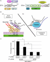
FIGURE 1.
FIGURE 1.
ZF21 is an adaptor protein that regulates disassembly of FAs and migration of cells. A, schematic illustration of the domain structures of ZF21 (left) and EEA1 (right). Binding proteins and regions of the respective proteins that interact with them are indicated. B, ZF21 resides in FAs (a) and on cytoplasmic vesicles (b). ZF21 binds to FAK and vesicles via the N-terminal FYVE domain. C, effect of FAK on ZF21-dependent cell migration was analyzed using MDA-MB-231 cells. Expression of either or both ZF21 and FAK was carried out by expressing the corresponding shRNAs. Knock down efficiency is presented under supplemental Fig. S2. Cells were subjected to a migration assay using a transwell chamber equipped with filters coated with fibronectin, and FBS was used as the attractant. First lane, shLacZ + shLacZ; second lane, shZF21 + shLacZ; third lane, shLacZ + shFAK; fourth lane, shZF21 + shFAK. Error bars represent the mean ± S.D. (n = 3). *, p < 0.005 (Student's t test).
FIGURE 2.
FIGURE 2.
The FYVE domain of ZF21 binds specifically to FAK. A, sequence alignment of the FYVE domains of ZF21 and EEA1. Conserved residues are denoted with asterisks. B, schematic representation of the GST fusion proteins used for the FAK pulldown assay. GST was fused at the N terminus of wild-type ZF21 (GST-WT), the FYVE domain of ZF21 (GST-ZF21FYVE), the FYVE domain of EEA1 (GST-EEA1FYVE), or two full-length ZF21 that has had its FYVE domain substituted by that of EEA1 (GST-SWAP). The numbers in the figure represent amino acid positions. C, whole cell lysates of HeLa cells were incubated with GST-tagged ZF21 derivatives or GST alone. Proteins bound to the GST fusion proteins were analyzed by Western blot using anti-FAK or anti-GST antibodies.
FIGURE 3.
FIGURE 3.
Analysis of the contribution of regions of the ZF21 protein required for FA disassembly and promotion of cell migration. A, schematic representation of the structures of the m1Venus-tagged fusion proteins of ZF21 expressed in ZF21-depleted MDA-MB231 cells (MDA-MB231(shZF21)). m1Venus was fused to the N terminus of wild-type ZF21 (m1V-WT), full-length ZF21 that has had its FYVE domain substituted by that of EEA1 (m1V-SWAP), or a ZF21 mutant lacking the C-terminal aa 106–234 region (m1V-N). B, each construct was stably expressed in MDA-MB231(shZF21) cells using a lentivirus vector system. The expression of each m1Venus-tagged protein was analyzed by Western blot using the indicated antibodies. The control (mock) represents cells infected with the empty lentiviral vector. C, immunostaining of MDA-MB-231(shZF21) cells for phospho-Tyr397-FAK. MDA-MB231(shZF21) cells were seeded onto fibronectin-coated glass coverslips and cultured for 48 h. The cells were treated with nocodazole (5 μm) for 30 min. Then the cells were washed and cultured with nocodazole-free media for 0 (a–g) or 30 min (h–n). The cells were subjected to immunostaining for phospho-Tyr397-FAK (red, a–d and h–k). ZF21 or the mutant thereof was visualized using the fluorescence of the tagged m1Venus protein (green, e–g and l–n). Scale bar, 20 μm. D, the same set of cells used in C was subjected to Western blot analysis using specific antibodies to detect the indicated forms of FAK. E, the cells were subjected to a migration assay using a transwell chamber equipped with filters coated with fibronectin and FBS was used as an attractant. First lane, shLacZ alone; second lane, shZF21 alone; third lane, shZF21 + mock vector; fourth lane, shZF21 + m1V-WT; fifth lane, shZF21 + m1V-SWAP; sixth lane, shZF21 + m1V-N. Error bars indicate the mean ± S.D. (n = 3). *, p < 0.005 (Student's t test).
FIGURE 4.
FIGURE 4.
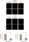
ZF21 promotes an early step of metastasis of tumor cells to the lung. As baseed on the reported methods (44, 45), metastasis of human tumor cells to the lung was analyzed by injecting the cells into the tail vein of mice. MDA-MB231 (A) or HT1080 (B) cells were used for the assay. Control cells were labeled with cell tracker orange and ZF21-depleted cells with cell tracker green. An equal number of the control and depleted cells were mixed and injected into the tail vein. The lung was dissected 1 or 24 h after injection and tumor cells in the tissue section were visualized (A and B) and the number of the cells counted (C). Error bars indicate the mean ± S.D. (n = 3). *, p < 0.005 (Student's t test).
FIGURE 5.
FIGURE 5.
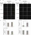
Analysis of the contribution of the FYVE and the C-terminal domains to the activity of ZF21 in the experimental metastasis assay. MDA-MB231 cells expressing shZF21 alone were labeled with red cell tracker and the cells expressing both shZF21 and each of the fusions shown in Fig. 3 were labeled with green cell tracker. A similar number of cells labeled with each color were mixed and analyzed as described in the legend to Fig. 4. Tissue sections analyzed at 1 (A) or 24 h (B) after injection are presented. The number of colored cells was counted and is presented in C. Error bars indicate the mean ± S.D. (n = 3). *, p < 0.005 (Student's t test).
FIGURE 6.
FIGURE 6.
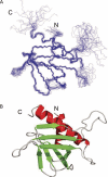
Structures of the C-terminal domain of ZF21. A, the ensemble of the final 20 best structures of the ZF21 C-terminal domain. The N and C termini are indicated as N and C, respectively. B, ribbon representation of the C-terminal domain is presented using a colored ribbon. The α-helices and β-strands are shown in red and green, respectively. The orientation is the same as in A.
FIGURE 7.
FIGURE 7.
Comparison of the structures of the ZF21 C-terminal and PLC-δ1 PH domains. A, the ZF21 C-terminal domain. B, crystal structure of the PLC-δ1 PH domain in complex with Ins(1,4,5)P3 (PDB code 1MAI). Left panels are ribbon representations. The α-helices and β-strands are shown in red and green, respectively. Ins(1,4,5)P3 is shown as a stick model. Right panels are electrostatic potential surfaces. The electrostatic potentials were calculated with the GRASP program (58). The range of the potential is from −5.0 (red) to +5.0 kT/e (blue). The orientations are the same as in the left panels.
FIGURE 8.
FIGURE 8.
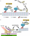
Possible function of the PH-like domain of ZF21. A, the PH-like domain of ZF21 may facilitate the loading of ZF21 onto transport vesicles localized to MTs and facilitates to convey the FA disassembly factors. Interaction of ZF21 with MTs via the PH-like domain and β-tubulin may represent an initial step of this process (i). ZF21 on MTs is thought to bind phosphoinositides within the transport vesicles (ii). ZF21 on MTs recruits FA disassembly factors, such as SHP-2 or m-calpain, to the transport vesicles (iii). The plus or minus end of MTs is indicated by symbols (+ or −), respectively. B, the PH-like domain of ZF21 may act as an acceptor site for MTs on FAs. MTs extended to FAs may be anchored to FAs by interacting with the PH-like domain of the ZF21 protein that already is localized to the FAs (a). ZF21 may recruit the heterodimeric α- and β-tubulin complex via the PH-like domain so as to provide substrates for the polymerization of MTs (b).

References

    1. Avraamides C. J., Garmy-Susini B., Varner J. A. (2008) Nat. Rev. Cancer 8, 604–617 - PMC - PubMed
    1. Desgrosellier J. S., Cheresh D. A. (2010) Nat. Rev. Cancer 10, 9–22 - PMC - PubMed
    1. Meighan C. M., Schwarzbauer J. E. (2008) Curr. Opin. Cell Biol. 20, 520–524 - PMC - PubMed
    1. Truong H., Danen E. H. (2009) Cell Adh. Migr. 3, 179–181 - PMC - PubMed
    1. Hynes R. O. (2002) Cell 110, 673–687 - PubMed

Publication types