. 2011 Sep;79(9):3596-606.
doi: 10.1128/IAI.00077-11. Epub 2011 Jul 18.

Mutations of the Listeria monocytogenes peptidoglycan N-deacetylase and O-acetylase result in enhanced lysozyme sensitivity, bacteriolysis, and hyperinduction of innate immune pathways

Affiliations

Mutations of the Listeria monocytogenes peptidoglycan N-deacetylase and O-acetylase result in enhanced lysozyme sensitivity, bacteriolysis, and hyperinduction of innate immune pathways

Chris S Rae et al. Infect Immun. 2011 Sep.

Abstract

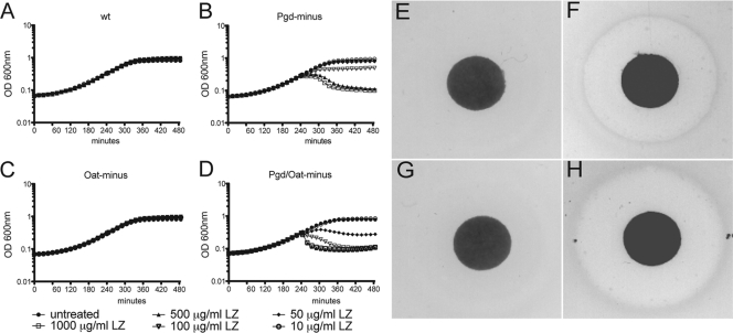
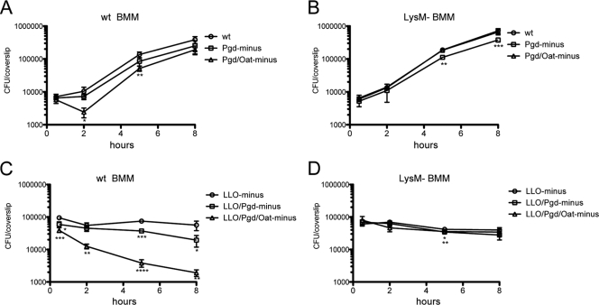
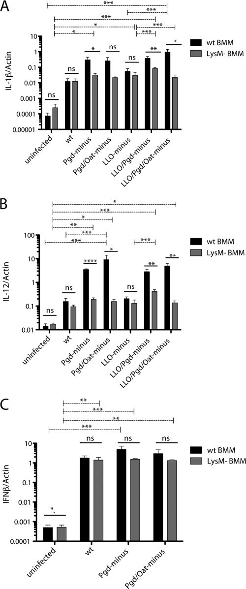
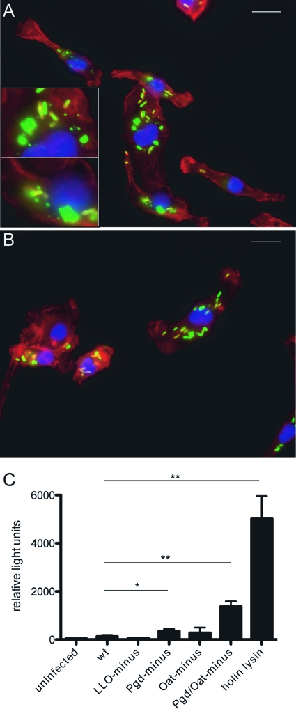
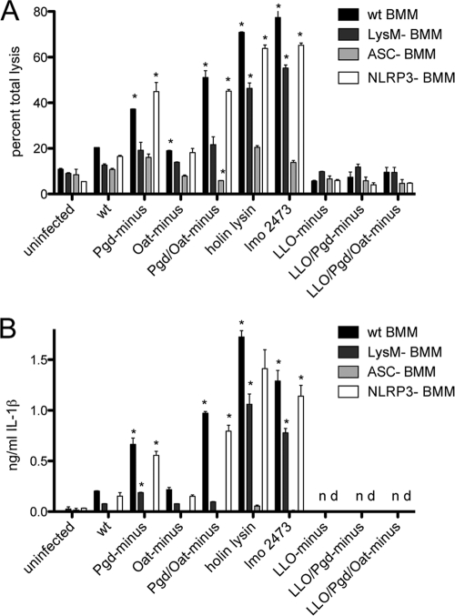
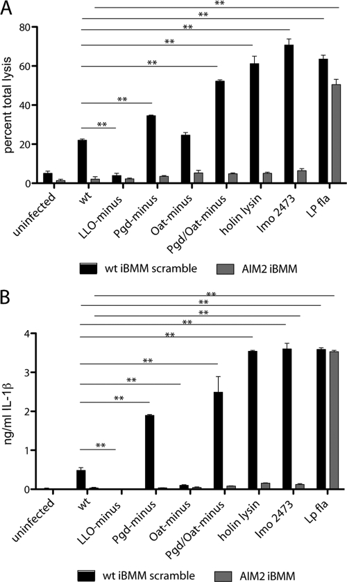
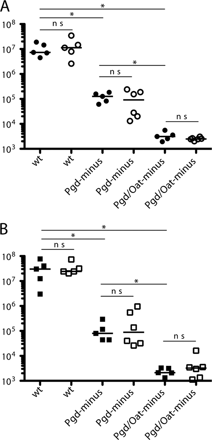
Listeria monocytogenes is a Gram-positive intracellular pathogen that is naturally resistant to lysozyme. Recently, it was shown that peptidoglycan modification by N-deacetylation or O-acetylation confers resistance to lysozyme in various Gram-positive bacteria, including L. monocytogenes. L. monocytogenes peptidoglycan is deacetylated by the action of N-acetylglucosamine deacetylase (Pgd) and acetylated by O-acetylmuramic acid transferase (Oat). We characterized Pgd(-), Oat(-), and double mutants to determine the specific role of L. monocytogenes peptidoglycan acetylation in conferring lysozyme sensitivity during infection of macrophages and mice. Pgd(-) and Pgd(-) Oat(-) double mutants were attenuated approximately 2 and 3.5 logs, respectively, in vivo. In bone-marrow derived macrophages, the mutants demonstrated intracellular growth defects and increased induction of cytokine transcriptional responses that emanated from a phagosome and the cytosol. Lysozyme-sensitive mutants underwent bacteriolysis in the macrophage cytosol, resulting in AIM2-dependent pyroptosis. Each of the in vitro phenotypes was rescued upon infection of LysM(-) macrophages. The addition of extracellular lysozyme to LysM(-) macrophages restored cytokine induction, host cell death, and L. monocytogenes growth inhibition. This surprising observation suggests that extracellular lysozyme can access the macrophage cytosol and act on intracellular lysozyme-sensitive bacteria.


Figures

Fig. 1.
Fig. 1.
Sensitivity of L. monocytogenes strains to lysozyme. Growth of wt (A), Pgd (B), Oat (C), and Pgd Oat (D) L. monocytogenes strains in BHI medium in the presence of titrated lysozyme (LZ). Lysozyme was added to cultures in equal volumes at 240 min after bacterial inoculation, and OD600 was monitored at 15-min intervals. Growth of wt (E), Pgd (F), Oat (G), and Pgd Oat (H) L. monocytogenes strains on LB plates containing 1 mg lysozyme on a Whatman paper disc. Bacterial lawns were grown overnight, and the zone of clearance was measured the next day. Data are representative of more than 3 independent experiments with similar results.
Fig. 2.
Fig. 2.
Growth of L. monocytogenes strains in BMM. A total of 2 × 10⁶ wt (A) or LysM (B) BMM were infected with wt, Pgd, or Pgd Oat L. monocytogenes at an MOI of 0.1. A total of 2 × 10⁶ wt (C) or LysM (D) BMM were infected with LLO L. monocytogenes strains at an MOI of 1. BMM were lysed, and bacterial CFU were quantified. Error bars represent standard deviations of the means of technical triplicates, and data are representative of more than 3 independent experiments with similar results. Student's t test was used to analyze statistical significance compared to wt L. monocytogenes infection at each time point. *, P < 0.05; **, P < 0.01; ***, P < 0.001; ****, P < 0.0001.
Fig. 3.
Fig. 3.
Pgd and Pgd Oat L. monocytogenes strains induce increased cytosolic and vacuolar cytokine responses dependent on the presence of host lysozyme. A total of 106 wt (black bars) or LysM (gray bars) BMM were infected with wt or lysozyme-sensitive L. monocytogenes at an MOI of 1 for 4 h. LLO infections were also performed at an MOI of 1. RNA was harvested, and IL-1β (A), IL-12 (B), and IFN-β (C) transcripts were measured relative to those of β-actin by quantitative PCR. Error bars represent standard deviations of the means determined in triplicate. Data are representative of at least 3 independent experiments with similar results. One-way analysis of variance (ANOVA) and Tukey's post hoc test were used to analyze the significance of infections in either wt or LysM BMM (dashed lines). Student's t test was used to analyze significance for each bacterial strain. *, P < 0.05; **, P < 0.01; ***, P < 0.001; ****, P < 0.0001; ns, not significant.
Fig. 4.
Fig. 4.
Intracellular bacteriolysis of lysozyme-sensitive L. monocytogenes. Immunofluorescence microscopy of wt (A) or LysM (B) BMM infected with Pgd Oat L. monocytogenes (green) imaged at 90 min postinfection (actin and nuclei were stained red and blue, respectively). Insets depict bacterial degradation. Size bars represent 10 μm. Cytosolic DNA delivery is increased in Pgd and Pgd Oat L. monocytogenes strains (C). IFN-α/βR BMM were infected with L. monocytogenes bearing a plasmid-containing luciferase under a CMV promoter at an MOI of 5. Luciferase expression was detected at 6 h postinfection. Error bars represent standard deviations of the means determined in triplicate. Statistical significance was evaluated using Student's t test. Data are representative of at least 3 separate experiments with similar results. *, P < 0.01; **, P < 0.0001.
Fig. 5.
Fig. 5.
Pgd and Pgd Oat L. monocytogenes strains induce increased inflammasome activation. (A) LDH release in wt, LysM, ASC, or NLRP3 Pam3CSK4-stimulated BMM. A total of 5 × 10⁵ BMM were infected for 6 h at an MOI of 5. (B) IL-1β secretion detected by ELISA in wt, LysM, ASC, or NLRP3 Pam3CSK4-stimulated BMM at 6 h postinfection at an MOI of 5. Error bars represent standard deviations of the means. Statistical significance was evaluated compared to wt L. monocytogenes infection using Student's t test. Data are representative of more than 3 independent experiments with similar results. *, P < 0.01.
Fig. 6.
Fig. 6.
Pgd and Pgd Oat L. monocytogenes strains activate the AIM2 inflammasome. (A) Pam3CSK4-stimulated immortalized shRNA AIM2 knockdown or shRNA scrambled macrophages were infected at an MOI of 5, and LDH was measured at 6 h. (B) IL-1β secretion was measured by ELISA in Pam3CSK4-stimulated immortalized shRNA AIM2 knockdown or shRNA scrambled immortalized macrophages (iBMM) infected for 6 h. Error bars represent standard deviations of the means determined in triplicate. Statistical significance was evaluated using Student's t test. Data are representative of more than 3 independent experiments with similar results. **, P < 0.0001. Lp fla, Legionella flagellin.
Fig. 7.
Fig. 7.
Bacterial replication but not cell-to-cell spread is required for release of LDH. LDH release over time is shown. (A) Pam3CSK4-stimulated wt BMM were infected at an MOI of 5, and LDH release was assayed at 3, 4.5, and 6 h postinfection. (B) A total of 0.1 mg/ml chloramphenicol (cm) was added at 1, 2, 3, 4, or 5 h postinfection, and LDH release was measured at 6 h postinfection. Error bars represent standard deviations of the means. Data are representative of more than 3 independent experiments with similar results.
Fig. 8.
Fig. 8.
Extracellular lysozyme accesses the BMM cytosol and results in increased degradation of lysozyme-sensitive L. monocytogenes strains and subsequent host cell death. (A) IFN-α/βR BMM were infected with L. monocytogenes bearing a plasmid-containing luciferase under a CMV promoter, and 1 mg/ml extracellular lysozyme was added at 1 h postinfection. Luciferase expression was detected at 6 h postinfection. (B) Pam3CSK4-stimulated LysM BMM were infected at an MOI of 5, and 1 mg/ml extracellular lysozyme was added at 1 h postinfection. LDH release was measured at 6 h. (C) Pam3CSK4-stimulated LysM BMM were treated with cycloheximide and infected at an MOI of 5, and LDH release was measured at 6 h. Error bars represent standard deviations of the means. (A and B) Statistical significance was evaluated using Student's t test. *, P < 0.05; ***, P < 0.0001. (C) One-way ANOVA was performed to analyze the statistical significance of LDH release following various treatments of BMM infected with individual L. monocytogenes strains. *, P < 0.05; **, P < 0.001. The means of LDH values from infections treated with lysozyme compared to those treated with lysozyme plus cycloheximide were determined to be not significant (ns) using Student's t test. The means of LDH values from infections with Pgd, Oat, or Pgd Oat L. monocytogenes were significant by one-way ANOVA. Data are representative of at least 3 independent experiments with similar results. LysM BMM were infected with either wt (D, E) or Pgd Oat (F, G) L. monocytogenes for 2 h. A total of 1 mg/ml extracellular lysozyme was added at 1 h postinfection (E, G), and cells were scored for evidence of bacterial degradation (depicted in inset). Size bars represent 10 μm. Data are representative of more than 3 independent experiments with similar results.
Fig. 9.
Fig. 9.
Pgd and Pgd Oat L. monocytogenes strains are defective in vivo but are not rescued in LysM mice. C57BL/6 or LysM mice were infected with 1 × 10⁵ bacteria by i.v. injection, and CFU were quantified in the spleen (A) and liver (B) at 48 h postinfection. Spleen and liver are represented by circles and squares, respectively. Filled and open shapes represent C57BL/6 and LysM mice, respectively. Statistical significance was evaluated using a Mann-Whitney test. Data are representative of more than 3 independent experiments with similar results. *, P < 0.02; ns, not significant.

References

    1. Bera A., Biswas R., Herbert S., Gotz F. 2006. The presence of peptidoglycan O-acetyltransferase in various staphylococcal species correlates with lysozyme resistance and pathogenicity. Infect. Immun. 74:4598–4604 - PMC - PubMed
    1. Bera A., Herbert S., Jakob A., Vollmer W., Gotz F. 2005. Why are pathogenic staphylococci so lysozyme resistant? The peptidoglycan O-acetyltransferase OatA is the major determinant for lysozyme resistance of Staphylococcus aureus. Mol. Microbiol. 55:778–787 - PubMed
    1. Boneca I. G., et al. 2007. A critical role for peptidoglycan N-deacetylation in Listeria evasion from the host innate immune system. Proc. Natl. Acad. Sci. U. S. A. 104:997–1002 - PMC - PubMed
    1. Callewaert L., Michiels C. W. 2010. Lysozymes in the animal kingdom. J. Biosci. 35:127–160 - PubMed
    1. Camilli A., Tilney L. G., Portnoy D. A. 1993. Dual roles of plcA in Listeria monocytogenes pathogenesis. Mol. Microbiol. 8:143–157 - PMC - PubMed

Publication types

MeSH terms

LinkOut - more resources