. 2011 Jul 25;194(2):177-86.
doi: 10.1083/jcb.201101062. Epub 2011 Jul 18.

APC/CCdh1-dependent proteolysis of USP1 regulates the response to UV-mediated DNA damage

Affiliations

APC/CCdh1-dependent proteolysis of USP1 regulates the response to UV-mediated DNA damage

Xiomaris M Cotto-Rios et al. J Cell Biol. .

Abstract

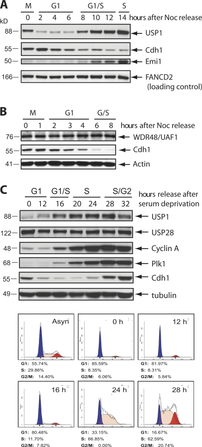
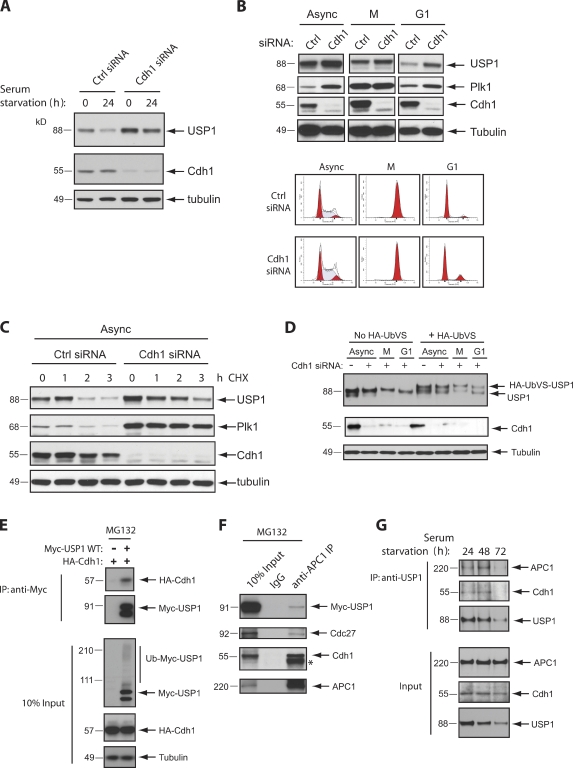
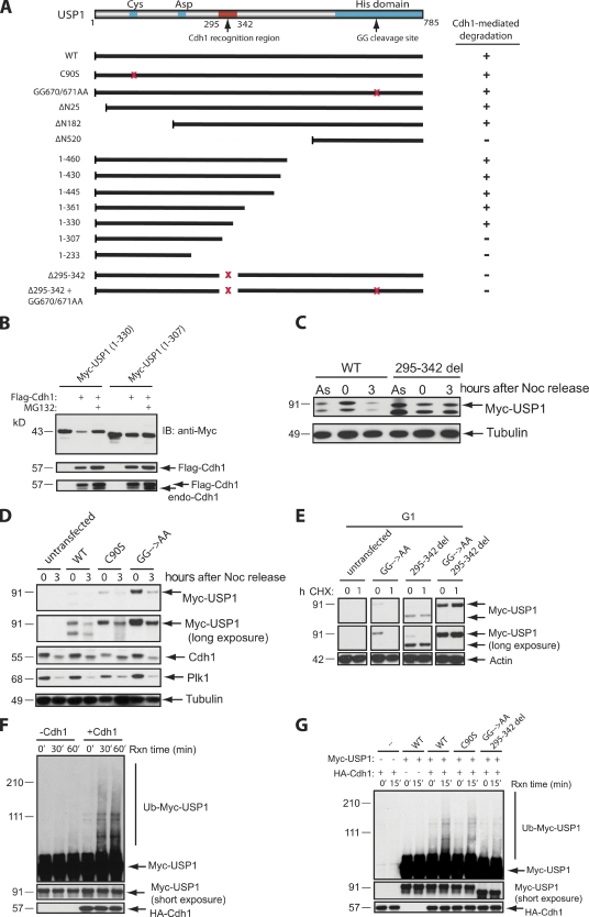
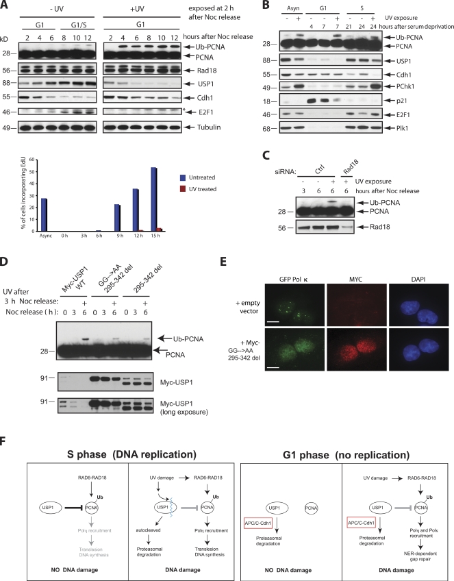
Targeted protein destruction of critical cellular regulators during the G1 phase of the cell cycle is achieved by anaphase-promoting complex/cyclosome(Cdh1) (APC/C(Cdh1)), a multisubunit E3 ubiquitin ligase. Cells lacking Cdh1 have been shown to accumulate deoxyribonucleic acid (DNA) damage, suggesting that it may play a previously unrecognized role in maintaining genomic stability. The ubiquitin-specific protease 1 (USP1) is a known critical regulator of DNA repair and genomic stability. In this paper, we report that USP1 was degraded in G1 via APC/C(Cdh1). USP1 levels were kept low in G1 to provide a permissive condition for inducing proliferating cell nuclear antigen (PCNA) monoubiquitination in response to ultraviolet (UV) damage before DNA replication. Importantly, expression of a USP1 mutant that cannot be degraded via APC/C(Cdh1) inhibited PCNA monoubiquitination during G1, likely compromising the recruitment of trans-lesion synthesis polymerase to UV repair sites. Thus, we propose a role for APC/C(Cdh1) in modulating the status of PCNA monoubiquitination and UV DNA repair before S phase entry.


Figures

Figure 1.
Figure 1.
USP1 levels are low during the G1 phase of the cell cycle. (A and B) HeLa (A) and U2OS (B) cells were synchronized in M phase by treating cells with nocodazole (Noc) for 16 h, washed, released, and collected for the indicated time points. (C) T98G human glioblastoma cells were synchronized in G0/G1 by serum deprivation for 72 h and then refed with serum and harvested at the indicated time points. (A–C) Western blot analyses were performed and probed with the indicated antibodies. Separate samples were also collected, fixed, and stained with PI for FACS analysis according to procedures outlined in the Materials and methods section.
Figure 2.
Figure 2.
Cdh1 depletion stabilizes USP1 during the G1 phase of the cell cycle. (A) T98G cells were transfected for 48 h with a control (Ctrl) siRNA (AllStars Negative; QIAGEN) or Cdh1 siRNA. Cells were serum starved in culture media containing 0.05% FBS for 24 h to arrest them in G0/G1. 0 indicates cells grown in regular media. (B) U2OS cells were transfected with the indicated siRNAs, synchronized in M phase (M) by incubating with nocodazole for 16 h, washed, and released in fresh media for 3 h for G1 phase (G1). Separate samples were collected for FACS. (C) U2OS cells were transfected with the indicated siRNAs and treated for the indicated time points with cycloheximide (CHX) to inhibit protein synthesis. (D) U2OS cells were transfected with the indicated siRNAs and synchronized in G1 as in B. Samples were collected and lysed according to protocols described for the UbVS DUB activity assay (see Materials and methods). Higher shift in the USP1 protein band indicates active USP1 (covalently modified USP1 by HA-UbVS). (E) Expression constructs were cotransfected in U2OS cells and treated for 6 h with 10 µM of the proteasome inhibitor MG132 before termination. Samples were lysed and collected for immunoprecipitation (IP) or loaded for input. (F) U2OS cells were transfected with Myc-USP1 wild type (WT) for 48 h and treated with MG132 as in E. Samples were lysed and collected for immunoprecipitation. *, heavy chain band. (G) T98G cells were serum starved as in A for the indicated times. Samples were lysed and collected for immunoprecipitation and input (10%) and probed with the indicated antibodies for Western blot analysis.
Figure 3.
Figure 3.
Identification of a USP1 mutant that cannot be degraded via APC/CCdh1. (A) A schematic diagram of critical USP1 domains and Myc-USP1 expression constructs generated for testing of their stability when coexpressed with FLAG-Cdh1. +, degradation of Myc-USP1; −, stabilization of Myc-USP1; GG, diglycine residue for the USP1 autocleavage site; red x’s, deletion or point mutations of the region. (B) U2OS cells were transiently transfected with Myc-USP1 and/or Flag-Cdh1 constructs in the presence or absence of MG132 (3 h). IB, immunoblot. (C) U2OS cells were transfected with Myc-USP1 wild type (WT) or the 295–342 deletion (295–342 del) mutant and synchronized in M phase with nocodazole (Noc) for 16 h (0 h) and/or released into G1 (3 h after nocodazole release). As, asynchronous. (D) U2OS cells were transfected with Myc-USP1 wild type, C90S, or GG670/671AA and synchronized in M phase or G1 as in C. (E) U2OS cells were transfected with Myc-USP1 wild type, GG670/671AA, 295–342 deletion, or GG670/671AA plus the 295–342 deletion mutant, synchronized, released into G1 (3 h after nocodazole release), and treated with cycloheximide (CHX) for 1 h (1) or not (0). (F) In vitro ubiquitination assay of Myc-USP1 wild type in the presence or absence of HA-Cdh1. Reactions were performed as described in the Materials and methods section. (G) In vitro ubiquitination assay of Myc-USP1 wild type, C90S, and GG670/671AA plus the 295–342 deletion mutant in the presence or absence of HA-Cdh1. Rxn, reaction.
Figure 4.
Figure 4.
UV-induced PCNA monoubiquitination during G1 requires prolonged degradation of USP1. (A) U2OS cells were synchronized in M phase with nocodazole (Noc) treatment and released for the indicated time points. Cells were also irradiated with 50 J/m2 UV in G1 (2 h after release from nocodazole treatment). Samples were then analyzed by Western blotting. Cells from corresponding samples were pulsed with EdU (BrdU analogue) to label cells undergoing DNA synthesis. Samples were processed according to procedures outlined in the Materials and methods section and were analyzed by FACS. The data displayed as a bar graph are representative of two separate experiments. (B) T98G cells were synchronized in G0/G1 by serum deprivation for 72 h, refed with fresh media, and collected at the indicated time points. Cells were either left untreated, UV exposed at 4 h after release from serum deprivation (G1 phase), or exposed after 21 h (early S phase). (C) U2OS cells were transfected with the indicated siRNAs, synchronized, released into G1 (3 h after nocodazole release), UV irradiated (50 J/m2), and collected 3 h after UV exposure (6 h after release). (D) U2OS cells were transfected with either Myc-USP1 wild type (WT), 295–342 deletion (295–342 del), or GG670/671AA plus the 295–342 deletion mutant, synchronized in M phase (0 h), released into G1 (3 h), UV irradiated (3 h after nocodazole release), and then collected 3 h after UV exposure (6 h). (E) U2OS cells stably expressing GFP–Pol-κ were selected in G418 for 10 d before isolation of GFP-positive cells through a FACS sorter (MoFlo; Dako). Stable U2OS GFP–Pol-κ–expressing cells were transiently transfected with Myc-tagged GG670/671AA plus the 295–342 deletion mutant, synchronized in M phase with nocodazole, and released for 3 h into the G1 phase. The cells were then irradiated with 50 J/m2 UV and fixed 2 h after UV exposure. Approximately 100 GFP-positive cells or GFP and Myc double-positive cells were analyzed. No Myc-expressing cells contained GFP–Pol-κ foci. A representative image of the cells analyzed is shown. GFP-positive nuclear foci formation was scored as the number of cells containing five or more foci. Bars, 20 µm. (F) A schematic representation of how USP1 is regulated by proteolysis in G1 versus S phase cells in the presence or absence of UV DNA damage. Black lines, active cells; gray lines, inactive cells; blue squiggly line, USP1 autocleavage site; Ub, ubiquitin.

References

    1. Aristarkhov A., Eytan E., Moghe A., Admon A., Hershko A., Ruderman J.V. 1996. E2-C, a cyclin-selective ubiquitin carrier protein required for the destruction of mitotic cyclins. Proc. Natl. Acad. Sci. USA. 93:4294–4299 10.1073/pnas.93.9.4294 - DOI - PMC - PubMed
    1. Bashir T., Dorrello N.V., Amador V., Guardavaccaro D., Pagano M. 2004. Control of the SCF(Skp2-Cks1) ubiquitin ligase by the APC/C(Cdh1) ubiquitin ligase. Nature. 428:190–193 10.1038/nature02330 - DOI - PubMed
    1. Bassermann F., Frescas D., Guardavaccaro D., Busino L., Peschiaroli A., Pagano M. 2008. The Cdc14B-Cdh1-Plk1 axis controls the G2 DNA-damage-response checkpoint. Cell. 134:256–267 10.1016/j.cell.2008.05.043 - DOI - PMC - PubMed
    1. Bienko M., Green C.M., Crosetto N., Rudolf F., Zapart G., Coull B., Kannouche P., Wider G., Peter M., Lehmann A.R., et al. 2005. Ubiquitin-binding domains in Y-family polymerases regulate translesion synthesis. Science. 310:1821–1824 10.1126/science.1120615 - DOI - PubMed
    1. Borodovsky A., Ovaa H., Kolli N., Gan-Erdene T., Wilkinson K.D., Ploegh H.L., Kessler B.M. 2002. Chemistry-based functional proteomics reveals novel members of the deubiquitinating enzyme family. Chem. Biol. 9:1149–1159 10.1016/S1074-5521(02)00248-X - DOI - PubMed

Publication types