. 2011 Sep 8;118(10):2793-800.
doi: 10.1182/blood-2011-01-331447. Epub 2011 Jul 18.

Therapeutic efficacy of FTY720 in a rat model of NK-cell leukemia

Affiliations

Therapeutic efficacy of FTY720 in a rat model of NK-cell leukemia

Aijun Liao et al. Blood. .

Abstract

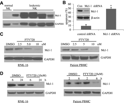
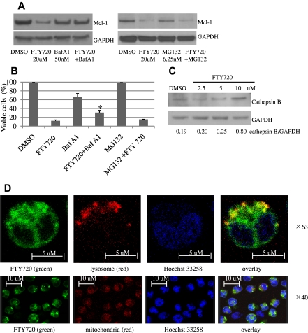
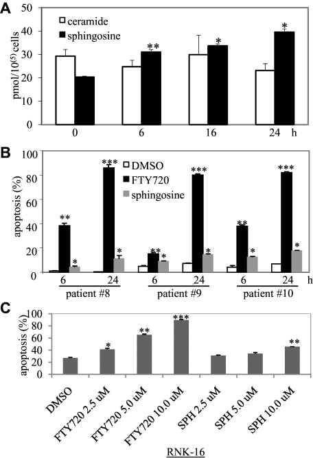
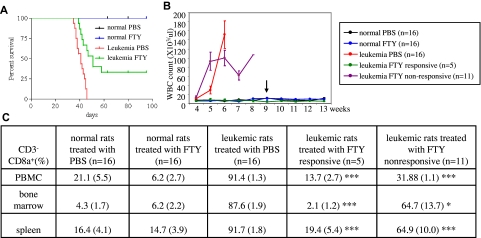
NK-cell leukemia is a clonal expansion of NK cells. The illness can occur in an aggressive or chronic form. We studied cell lines from human and rat NK-cell leukemias (aggressive NK-cell leukemia) as well as samples from patients with chronic NK-cell leukemia to investigate pathogenic mechanisms. Here we report that Mcl-1 was overexpressed in leukemic NK cells and that knockdown of Mcl-1 induced apoptosis in these leukemic cells. In vitro treatment of human and rat NK leukemia cells with FTY720 led to caspase-dependent apoptosis and decreased Mcl-1 expression in a time- and-dose-dependent manner. These biologic effects could be inhibited by blockade of reactive oxygen species generation and the lysosomal degradation pathway. Lipidomic analyses after FTY720 treatment demonstrated elevated levels of sphingosine, which mediated apoptosis of leukemic NK cells in vitro. Importantly, systemic administration of FTY720 induced complete remission in the syngeneic Fischer rat model of NK-cell leukemia. Therapeutic efficacy was associated with decreased expression of Mcl-1 in vivo. These data demonstrate that therapeutic benefit of FTY720 may result from both altered sphingolipid metabolism as well as enhanced degradation of a key component of survival signaling.


Figures

Figure 1
Figure 1
FTY720 induces apoptotic cell death in leukemic NK cells in a dose-dependent manner. (A) Human NKL cell line was treated with 5, 10, and 15μM FTY720 or DMSO for 24 hours, then cells were assayed for apoptosis by flow cytometry. (B) Rat RNK-16 cell line was treated with 2.5, 5, and 10μM FTY720 or DMSO for 24 hours then cells were assayed for apoptosis by flow cytometry. (C) PBMCs from one acute NK-cell leukemia patient (no. 1) and 3 chronic NK-cell leukemia patients (nos. 2, 3, 4) were treated with 2.5, 5, and 10μM FTY720 or DMSO for 18 hours, then cells were assayed for apoptosis by flow cytometry. *P < .05, **P < .005, and ***P < .0005 indicate significant differences of FTY720-treated cells versus DMSO-treated cells (Student t test).
Figure 2
Figure 2
Mcl-1 plays an essential role in survival of leukemic NK cells and is degraded by FTY720. (A) Increased expression of Mcl-1 in NK-cell leukemia. Proteins were isolated from PBMCs of 5 individual patients with chronic NK-cell leukemia (CD3CD56+ cells > 80%), or pooled enriched NK cells (CD3CD56+ 80%-95%) from 10 normal human donors, then resolved in the SDS-PAGE gel loading buffer in a boiling water bath for 5 minutes. Western blot analysis was performed for detection of Mcl-1. GAPDH was used as a loading control. (B) Knockdown of Mcl-1 induces apoptosis in leukemic NK cells. Human NKL cells were infected with the concentrated PLKO.1 Mcl-1 shRNA or scramble shRNA lentiviral stocks for 48 hours, and then apoptosis assay was performed. *P < .05 indicates significant difference in apoptosis of Mcl-1 shRNA-infected cells compared with control shRNA-infected cells (Student t test). (Inset) Western blot analysis was performed for Mcl-1 in the control shRNA or Mcl-1 shRNA-infected NKL cells 48 hours after infection. The equal loading of protein was confirmed by probing with GAPDH Ab. (C) Dose-dependent reduced expression of Mcl-1 after FTY720 treatment. Western blot analysis was performed for Mcl-1 after 24-hour treatment of RNK-16 cells or PBMC cells from an NK-cell leukemia patient, with 2.5, 5, and 10μM FTY720. Data are representative of 3 independent experiments on RNK-16 cells or PBMCs from 3 chronic NK-cell leukemia patients. (D) Time-dependent decreased expression of Mcl-1 after FTY720 treatment. Western blot analysis was performed for Mcl-1 after treatment of RNK-16 cells with 10μM FTY720 for 6 and 24 hours, or PBMC cells from an NK-cell leukemia patient with 5μM FTY720 for 2 and 6 hours. Data are representative of 3 independent experiments on RNK-16 cells or PBMCs from 3 chronic NK-cell leukemia patients.
Figure 3
Figure 3
FTY720 down-regulates Mcl-1 expression via lysosomal degradation. RNK-16 cells were pretreated with 50nM bafilomycin A1 or 6.25nM MG132 for 30 minutes then exposed to 20μM FTY720 for 3 hours, then Western blot was performed to examine Mcl-1 expression (A) or cells were assayed for cell viability by MTT (B). (C) Dose-dependent increased expression of cathepsin B after FTY720 treatment. Western blot analysis was performed for cathepsin B in cytosolic fraction of RNK-16 cells after 24-hour treatment with 2.5, 5, and 10μM FTY720. ImageJ software was used to analyze the ratio of densitometry of cathepsin B to GAPDH as labeled in the bottom of Western blot images. (D) Confocal microscopic image of RNK-16 cells after 2 hours of treatment, showing that yellow staining indicating NBD-FTY720 (green) colocalized with cellular lysosome (top panel, red, magnification: ×63) but not mitochondria (bottom panel, red, magnification: ×40). Cellular nucleus was stained with (Hoechst 33258 blue).
Figure 4
Figure 4
FTY720 increased production of apoptosis-inducing lipid sphingosine. (A) Five million RNK-16 cells were treated with 10μM FTY720 for 6, 16, and 24 hours then were extracted to obtain lipids; extracts were then identified and quantitated by tandem mass spectrometry. Each sample had 4 replicates. *P < .05 and **P < .005 indicate significant differences versus control (Student t test). (B) PBMCs from 3 individual chronic NK-cell leukemia patients (nos. 8, 9, 10) were treated with 5μM FTY720, 5μM sphingosine, or DMSO for 6 and 24 hours then cells were assayed for apoptosis by flow cytometry. (C) RNK-16 cells were treated with 2.5, 5.0, 10.0μM FTY720 or sphinginosine (SPH) for 48 hours, then cells were assayed for apoptosis by flow cytometry. *P < .05, **P < .005, and ***P < .0005 indicate significant differences versus control (Student t test).
Figure 5
Figure 5
FTY720 induces complete remission in NK-cell leukemic rats. (A) The Kaplan-Meier survival curves for normal rats after treatment with 4.5 mg/kg of FTY720 (n=16) or PBS (n=16) or leukemic rats after treatment with either 4.5 mg/kg (n=16) or PBS (n=16), were plotted. (B) Maintenance of normal white blood counts, hemoglobin values, and platelet counts in responding leukemic and normal rats treated with FTY720. Blood (200 μL) from leukemic rats treated with FTY720 (n=16) and PBS (n=16) and normal rats treated with FTY720 (n=16) and PBS (n=16) was collected every week from tail veins of the animals and placed in EDTA K2-coated tubes, then CBC (complete blood count) analysis was performed. Arrow indicates the cessation of the treatment. (C) Flow cytometry was used to identify rat leukemic NK cells, which are CD3CD8a+. Comparison of CD3CD8a+ NK cells isolated from multiple tissues among normal rats treated with PBS (n=16) or FTY720 (n=16), leukemic rats treated with PBS (n=16) or leukemic rats responsive to FTY720 treatment (n=5) or unresponsive to FTY720 (n=11). *P < .05, **P < .005, ***P < .005 indicates significance between leukemic rats responsive to FTY720 or unresponsive to FTY20 and leukemic rats treated with PBS (unpaired t test).

References

    1. Lim MS, de Leval L, Quintanilla-Martinez L. Commentary on the 2008 WHO classification of mature T- and NK-cell neoplasms. J Hematop. 2009;2:75–76. - PMC - PubMed
    1. Lamy T, Loughran TP. Large granular lymphocyte leukemia. Cancer Control. 1998;5(1):25–33. - PubMed
    1. Sokol L, Loughran TP., Jr Large granular lymphocyte leukemia. Oncologist. 2006;11(3):263–273. - PubMed
    1. Cheung MM, Chan JK, Wong KF. Natural killer cell neoplasms: a distinctive group of highly aggressive lymphomas/leukemias. Semin Hematol. 2003;40(3):221–232. - PubMed
    1. Gold R. Oral therapies for multiple sclerosis: a review of agents in phase III development or recently approved. CNS Drugs. 2011;25(1):37–52. - PubMed

Publication types

MeSH terms