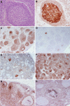
Follicular lymphoma in situ: clinical implications and comparisons with partial involvement by follicular lymphoma
- PMID: 21768298
- PMCID: PMC3175777
- DOI: 10.1182/blood-2011-05-355255
Follicular lymphoma in situ: clinical implications and comparisons with partial involvement by follicular lymphoma
Abstract
Follicular lymphoma in situ (FLIS) was first described nearly a decade ago, but its clinical significance remains uncertain. We reevaluated our original series and more recently diagnosed cases to develop criteria for the distinction of FLIS from partial involvement by follicular lymphoma (PFL). A total of 34 cases of FLIS were identified, most often as an incidental finding in a reactive lymph node. Six of 34 patients had prior or concurrent FL, and 5 of 34 had FLIS composite with another lymphoma. Of patients with negative staging at diagnosis and available follow-up (21 patients), only one (5%) developed FL (follow-up: median, 41 months; range, 10-118 months). Follow-up was not available in 2 cases. Fluorescence in situ hybridization for BCL2 gene rearrangement was positive in all 17 cases tested. PFL patients were more likely to develop FL, diagnosed in 9 of 17 (53%) who were untreated. Six patients with PFL were treated with local radiation therapy (4) or rituximab (2) and remained with no evidence of disease. FLIS can be reliably distinguished from PFL and has a very low rate of progression to clinically significant FL. FLIS may represent the tissue counterpart of circulating t(14;18)-positive B cells.
Figures


References
-
- Groves FD, Linet MS, Travis LB, Devesa SS. Cancer surveillance series: non-Hodgkin's lymphoma incidence by histologic subtype in the United States from 1978 through 1995. J Natl Cancer Inst. 2000;92(15):1240–1251. - PubMed
-
- Non-Hodgkin's Lymphoma Classification Project. A clinical evaluation of the International Lymphoma Study Group classification of non-Hodgkin's lymphoma. Blood. 1997;89(11):3909–3918. - PubMed
-
- Swerdlow SH, Campo E, Harris NL, et al. WHO Classification of Tumours of Haematopoietic and Lymphoid Tissues. 4th ed. Lyon, France: International Agency for Research on Cancer; 2008.
-
- Horning SJ, Rosenberg SA. The natural history of initially untreated low-grade non-Hodgkin's lymphomas. N Engl J Med. 1984;311(23):1471–1475. - PubMed
-
- Ardeshna KM, Smith P, Norton A, et al. Long-term effect of a watch and wait policy versus immediate systemic treatment for asymptomatic advanced-stage non-Hodgkin lymphoma: a randomised controlled trial. Lancet. 2003;362(9383):516–522. - PubMed
Publication types
MeSH terms
Substances
Grants and funding
LinkOut - more resources
Full Text Sources
Other Literature Sources
Research Materials

