. 2011 Aug 2;108(31):12723-8.
doi: 10.1073/pnas.1105128108. Epub 2011 Jul 18.

Reluctance to membrane binding enables accessibility of the synaptobrevin SNARE motif for SNARE complex formation

Affiliations

Reluctance to membrane binding enables accessibility of the synaptobrevin SNARE motif for SNARE complex formation

Kyle D Brewer et al. Proc Natl Acad Sci U S A. .

Abstract

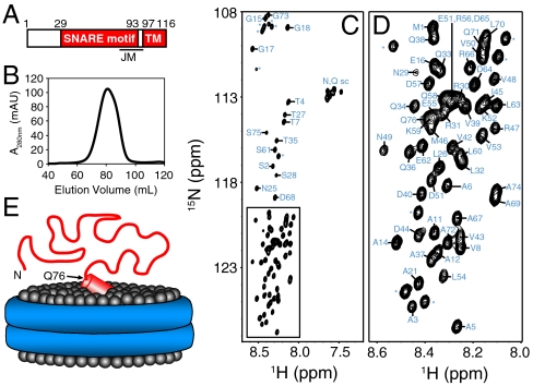
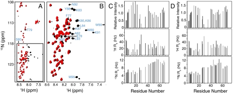
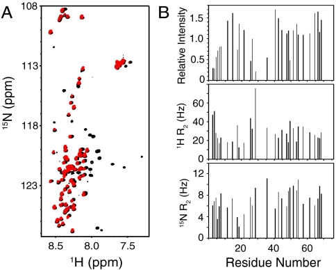
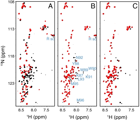
SNARE proteins play a critical role in intracellular membrane fusion by forming tight complexes that bring two membranes together and involve sequences called SNARE motifs. These motifs have a high tendency to form amphipathic coiled-coils that assemble into four-helix bundles, and often precede transmembrane regions. NMR studies in dodecylphosphocholine (DPC) micelles suggested that the N-terminal half of the SNARE motif from the neuronal SNARE synaptobrevin binds to membranes, which appeared to contradict previous biophysical studies of synaptobrevin in liposomes. NMR analyses of synaptobrevin reconstituted into nanodiscs and into liposomes now show that most of its SNARE motif, except for the basic C terminus, is highly flexible, exhibiting cross-peak patterns and transverse relaxation rates that are very similar to those observed in solution. Considering the proximity to the bilayer imposed by membrane anchoring, our data show that most of the synaptobrevin SNARE motif has a remarkable reluctance to bind membranes. This conclusion is further supported by NMR experiments showing that the soluble synaptobrevin SNARE motif does not bind to liposomes, even though it does bind to DPC micelles. These results show that nanodiscs provide a much better membrane model than DPC micelles in this system, and that most of the SNARE motif of membrane-anchored synaptobrevin is accessible for SNARE complex formation. We propose that the charge and hydrophobicity of SNARE motifs is optimized to enable formation of highly stable SNARE complexes while at the same time avoiding membrane binding, which could hinder SNARE complex assembly.


Conflict of interest statement

The authors declare no conflict of interest.

Figures

Fig. 1.
Fig. 1.
NMR analysis of synaptobrevin in nanodiscs. (A) Domain structure of synaptobrevin. Residue numbers above the bar indicate the sequence spanning the SNARE motif and the TM region. The approximate position of the juxtamembrane region (JM), which includes the C terminus of the SNARE motif and the linker joining it to the TM region, is indicated below. (B) Gel filtration on a Superdex200 column of the nanodiscs containing synaptobrevin after detergent removal and before concentrating for NMR analysis. (C, D) 1H-15N HSQC spectrum of synaptobrevin incorporated into nanodiscs. The expansion shown in D corresponds to the box of C. Cross-peak assignments are indicated (* indicates those from N-terminal residues arising from the expression vector). (E) Cartoon representing the overall structure of synaptobrevin (red) in nanodiscs with the lipid headgroups shown as gray spheres and the ApoA1 scaffold shown as a double blue ring. The diagram is meant to illustrate that residues 1–76 of synaptobrevin are highly flexible. The juxtamembrane region is represented by a tilted cylinder that represents a helix and is bound on the surface of the nanodiscs in a tilted orientation based on EPR data (25).
Fig. 2.
Fig. 2.
Residues 1–74 of synaptobrevin have a similar conformational behavior in solution and on nanodiscs. (A, B) Superposition of 1H-15N HSQC spectra of the soluble synaptobrevin(1–96) fragment (black contours) and full-length synaptobrevin in nanodiscs composed of POPC∶DOPS 85∶15 (red contours). The expansion shown in B corresponds to the box of A. Cross-peak assignments are indicated for residues from the soluble synaptobrevin(1–96) fragment that are not observed on nanodiscs. (C, D) Relative cross-peak intensities (Top), and transverse relaxation rates (R2) of the 1H (Middle) and 15N (Bottom) backbone nuclei of residues 1–74 of the soluble synaptobrevin(1–96) fragment (C) or of full-length synaptobrevin in nanodiscs (D). Data were quantified only for well-resolved cross-peaks. Relative intensities were calculated by dividing the observed intensity of a cross-peak by the average of all cross-peak intensities (46).
Fig. 3.
Fig. 3.
Residues 1–74 of synaptobrevin have a similar conformational behavior in solution and on liposomes. (A) Superposition of 1H-15N HSQC spectra of the soluble synaptobrevin(1–96) fragment (black contours) and full-length synaptobrevin reconstituted into liposomes composed of POPC∶DOPS 85∶15 (molar ratio) (red contours). (B) Relative cross-peak intensities (Top), and transverse relaxation rates (R2) of the 1H (Middle) and 15N (Bottom) backbone nuclei of residues 1–74 of full-length synaptobrevin in liposomes (same sample as A). The data were analyzed as in Fig. 2 C and D.
Fig. 4.
Fig. 4.
The N-terminal half of the SNARE motif of soluble synaptobrevin binds to DPC micelles but not to liposomes. (AC) Superposition of 1H-15N HSQC spectra of the 10 μM soluble synaptobrevin(1–96) fragment (black contours) alone or in the presence (red contours) of 300 mM DPC at pH 6.0 (A), POPC liposomes (50 mM lipids) at pH 6.0 (B), or POPC liposomes (50 mM lipids) at pH 7.0 (C). In B, cross-peaks that disappear upon addition of liposomes are labeled; cross-peaks from Arg side chains, which are observable at pH 6.0 but not at pH 7.0, are labeled Rsc. Note that in A there are less observable cross-peaks in DPC than in ref.  because we used a much lower protein concentration, but the data are fully consistent.

References

    1. Wickner W, Schekman R. Membrane fusion. Nat Struct Mol Biol. 2008;15:658–664. - PMC - PubMed
    1. Jahn R, Scheller RH. SNAREs—Engines for membrane fusion. Nat Rev Mol Cell Biol. 2006;7:631–643. - PubMed
    1. Brunger AT. Structure and function of SNARE and SNARE-interacting proteins. Q Rev Biophys. 2005;38:1–47. - PubMed
    1. Rizo J, Rosenmund C. Synaptic vesicle fusion. Nat Struct Mol Biol. 2008;15:665–674. - PMC - PubMed
    1. Sudhof TC, Rothman JE. Membrane fusion: Grappling with SNARE and SM proteins. Science. 2009;323:474–477. - PMC - PubMed

Publication types

MeSH terms

LinkOut - more resources