. 2011 Aug 2;108(31):12881-6.
doi: 10.1073/pnas.1101006108. Epub 2011 Jul 18.

Serine protease autotransporters from Shigella flexneri and pathogenic Escherichia coli target a broad range of leukocyte glycoproteins

Affiliations

Serine protease autotransporters from Shigella flexneri and pathogenic Escherichia coli target a broad range of leukocyte glycoproteins

Fernando Ruiz-Perez et al. Proc Natl Acad Sci U S A. .

Abstract

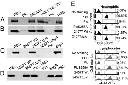
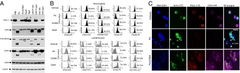
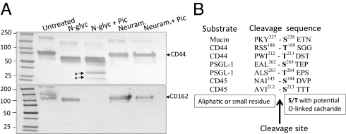
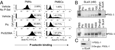
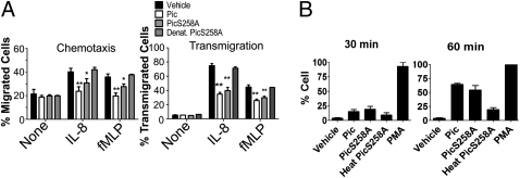
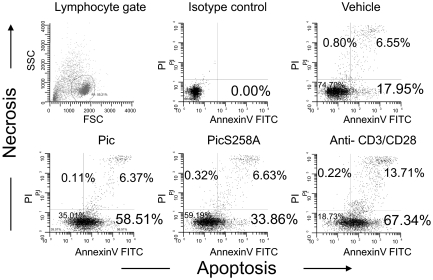
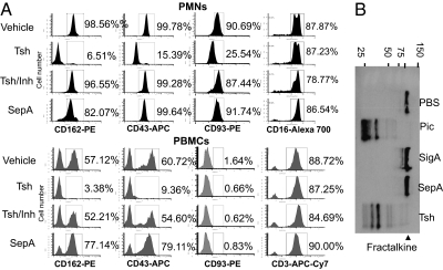
The serine protease autotransporters of Enterobacteriaceae (SPATEs) are secreted by pathogenic Gram-negative bacteria through the autotransporter pathway. We previously classified SPATE proteins into two classes: cytotoxic (class 1) and noncytotoxic (class 2). Here, we show that Pic, a class 2 SPATE protein produced by Shigella flexneri 2a, uropathogenic and enteroaggregative Escherichia coli strains, targets a broad range of human leukocyte adhesion proteins. Substrate specificity was restricted to glycoproteins rich in O-linked glycans, including CD43, CD44, CD45, CD93, CD162 (PSGL-1; P-selectin glycoprotein ligand 1), and the surface-attached chemokine fractalkine, all implicated in leukocyte trafficking, migration, and inflammation. N-terminal sequencing of proteolytic products revealed Pic (protease involved in colonization) cleavage sites to occur before Thr or Ser residues. The purified carbohydrate sLewis-X implied in inflammation and malignancy inhibited cleavage of PSGL-1 by Pic. Exposure of human leukocytes to purified Pic resulted in polymorphonuclear cell activation, but impaired chemotaxis and transmigration; Pic-treated T cells underwent programmed cell death. We also show that the Pic-related protease Tsh/Hbp, implicated in extraintestinal infections, exhibited a spectrum of substrates similar to those cleaved by Pic. In the guinea pig keratoconjunctivitis model, a Shigella pic mutant induced greater inflammation than its parent strain. We suggest that the class-2 SPATEs represent unique immune-modulating bacterial virulence factors.


Conflict of interest statement

The authors declare no conflict of interest.

Figures

Fig. 1.
Fig. 1.
Cleavage of sialomucin (CD43) on human leukocytes by Pic. PMNs (A, C, and E) and PBMCs (B, D, and E) were isolated from healthy volunteers and treated with log-phase supernatants from the following strains: EAEC 042, an isogenic EAEC042Δpic mutant, or the protease deficient EAEC042PicS258A strain; controls comprised 2 μM of purified Pic protein or PBS (A and B). Cells were alternatively treated with supernatants of S. flexneri 2457T, an isogenic Pic mutant, an isogenic SigA null mutant strain, or with 2 μM of purified Pic, PicS258A, or SigA (C and D). Following 30-min incubation at 37 °C, samples were analyzed by SDS/PAGE followed by immunoblot (A–D) or flow cytometry (E) using an Hrp- or APC-conjugated monoclonal antibody specific to the extracellular domain of human CD43. PBMC were gated by low side scatter, and PMNs were gated under high side scatter. Flow cytometry data are representative of at least four independent experiments.
Fig. 2.
Fig. 2.
Cleavage of an O-glycosylated mucin-like protein family by Pic. (A) Five micrograms of glycosylated human recombinant proteins were incubated at 37 °C for 1 h with supernatants of S. flexneri 2457T, EAEC 042, the isogenic S. flexneriΔpic and EAEC042Δpic mutant strains, or with 2 μM of purified Pic, PicS258A, SepA, or SigA. Samples were analyzed by SDS/PAGE followed by immunoblot using monoclonal antibodies to the external domain of PSGL-1, CD45, CD44, and fractalkine. Control proteins included ICAM-1 (CD54) and LAMP-1. (B) Pic protein degrades the extracellular domain of O-glycosylated mucin-like proteins on human leukocytes. The 1 × 10⁶ PMNs or PBMCs were isolated from human blood and incubated at 37 °C for 30 min with 2 μM of purified Pic, PicS258A, or SepA and analyzed by flow cytometry using monoclonal antibodies against the extracellular domains of CD44, CD45, PSGL-1, CD93, CD3, or CD16. Lymphocytes were gated by low side scatter using anti-CD3, but neutrophils were gated under high side scatter and anti-CD16 binding. Flow cytometry data are representative of at least four independent experiments. (C) To visualize degradation of mucin-like proteins, Pic-treated whole-blood leukocytes were stained for DNA, actin, CD43, and PSGL-1, followed by fluorescence microscopy. Arrows indicate PMNs and lymphocytes by virtue of nucleus and cytoplasm shape.
Fig. 3.
Fig. 3.
Proteolytic activity of Pic is dependent on O-, but not on N-glycosylation. (A) Five micrograms of recombinant CD44 or PSGL-1 proteins were de-glycosylated with neuraminidase or N-glycosydase-F at 37 °C overnight; deglycosylated proteins were then incubated with 2 μM of Pic at 37 °C for 1 h. Samples were analyzed by Western immunoblot using an anti-CD44 or anti-PSGL-1 antibodies. (B) Amino acid sequences flanking cleavage sites in four Pic substrates are shown. Asterisks indicate degraded products analyzed by N-terminal sequencing.
Fig. 4.
Fig. 4.
Inhibition of P-selectin binding to human leukocyte by Pic. (A) 1 × 10⁶ human lymphocytes were treated with 2 μM purified Pic or PicS258A, or with PBS control for 30 min followed for 30-min incubation with human P-selectin-IgG chimera. Binding of P-selectin chimera to leukocytes was evaluated by flow cytometry using a FITC-conjugated anti human Fc antibody. Untreated cells without incubation with P-selectin chimera (No P-Sel) were also stained with antihuman Fc antibody. (B) Inhibition of Pic protease activity by SLeX carbohydrate was evaluated by Western blot using 5 μg of recombinant PSGL-1 and 2 μM of Pic previously incubated with twofold increasing concentration of SLeX carbohydrate (5–1,600 nM). PSGL-1 or PSGL-1 deglycosylated by neuraminidase treatment (De-glyc) were incubated with Pic as controls. PBS, untreated PSGL-1; α-SLeX, PSGL-1 previously incubated with an anti-SLeX antibody before treatment with Pic. Western blot was developed with an anti PSGL-1 monoclonal antibody. (C) SLeX saccharide on PSGL-1 and deglycosylated PSGL-1 were analyzed by Western blot using a monoclonal antibody against SLeX.
Fig. 5.
Fig. 5.
Impairment of PMN chemotaxis and transmigration through endothelial cell monolayers by Pic. (A) Chemotaxis: 3 × 10⁵ calcein-AM labeled PMNs were treated with 2 μM of Pic, PicS258A, or PBS vehicle control in the upper chamber of Fbg-coated transwell inserts; 100 mM of IL-8 or 100 nM of fMLP were added to the lower chamber. Penetration of cells through the membrane was measured after 4 h fluorometrically. Transmigration: PMNs preincubated as above were applied to the upper chamber of inserts supporting HBMVEC-L, and transmigrated PMNs were enumerated as mentioned before. Data shown are means and SEM of at least three independent experiments performed in triplicate; dates were combined and analyzed by one-way ANOVA. The numbers of migrated cells were normalized to vehicle control with chemoattractant (**P < 0.01, *P < 0.05). (B) Activation of neutrophil oxidative burst by Pic: 1 × 10⁶ human neutrophils were labeled with dihydrorhodamine-123 dye for 5 min, immediately treated with 2 μM of LPS-free purified Pic, PicS258A, heat-denatured PicS258A, or PBS vehicle control, and analyzed by flow cytometry for oxidative burst, indicated by FITC fluorescence at 30- and 60-min time points. For positive control, 100 ng/mL of PMA was used. The cell population was gated to distinguish vehicle-treated and PMA-treated controls. Flow cytometry data are representative of three independent experiments.
Fig. 6.
Fig. 6.
Pic triggers cell death of activated human T cells. Activated human T lymphocytes on day 5 after stimulation with ConA (2 μg/mL) were incubated with 2 μM of purified Pic, PicS258A, or control anti-CD3/CD28 antibodies overnight. Following double staining with annexin-V FITC and PI, cells were analyzed by flow cytometry to determine the percentage of dead cells. Without gating, 10% of 10,000 total events were displayed. Flow cytometry data are representative of at least three independent experiments.
Fig. 7.
Fig. 7.
Tsh degrades the extracellular domain of O-glycosylated mucin-like proteins on human leukocytes. (A) 1 × 10⁶ PMNs and PBMCs were isolated from human blood and incubated at 37 °C for 30 min. with 1 μM of purified Tsh or Tsh previously inactivated with AEBSF (serine protease inhibitor). SepA was used as a protein control for digestion. Samples were analyzed by flow cytometry using monoclonal antibodies against the extracellular domain of PSGL-1, CD43, CD93, CD3, and CD16. PMNs and PBMCs were gated as in Fig 2B. Flow cytometry results are representative of at least three independent experiments. (B) Human recombinant fractalkine was treated with 2 μM of purified Pic, SepA, Tsh, or SigA for 1 h at 37 °C and analyzed by Western blot using a monoclonal antibody against human fractalkine.

References

    1. Kaper JB, Nataro JP, Mobley HL. Pathogenic Escherichia coli. Nat Rev Microbiol. 2004;2:123–140. - PubMed
    1. O'Ryan M, Prado V, Pickering LK. A millennium update on pediatric diarrheal illness in the developing world. Semin Pediatr Infect Dis. 2005;16:125–136. - PubMed
    1. Henderson IR, Navarro-Garcia F, Nataro JP. The great escape: Structure and function of the autotransporter proteins. Trends Microbiol. 1998;6:370–378. - PubMed
    1. Henderson IR, Nataro JP. Virulence functions of autotransporter proteins. Infect Immun. 2001;69:1231–1243. - PMC - PubMed
    1. Dutta PR, Cappello R, Navarro-García F, Nataro JP. Functional comparison of serine protease autotransporters of enterobacteriaceae. Infect Immun. 2002;70:7105–7113. - PMC - PubMed

Publication types

MeSH terms