. 2011 Aug 2;108(31):12839-44.
doi: 10.1073/pnas.1102167108. Epub 2011 Jul 18.

Structural basis for complement factor I control and its disease-associated sequence polymorphisms

Affiliations

Structural basis for complement factor I control and its disease-associated sequence polymorphisms

Pietro Roversi et al. Proc Natl Acad Sci U S A. .

Abstract

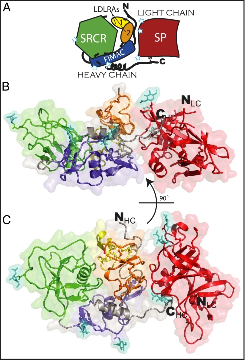
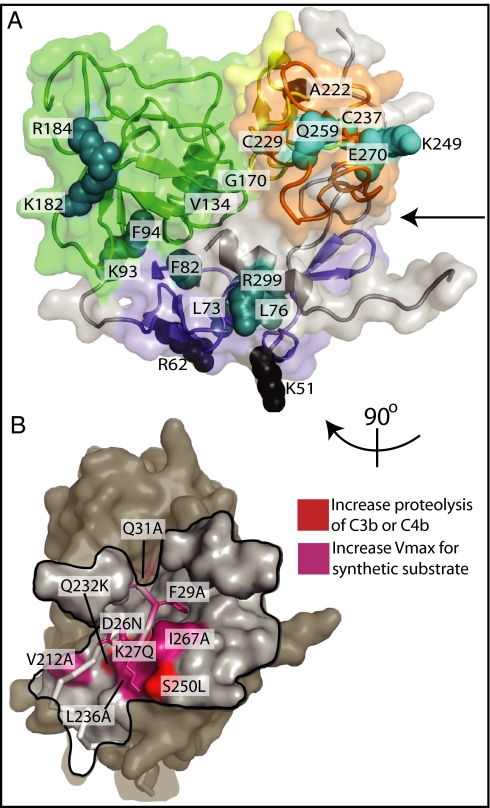
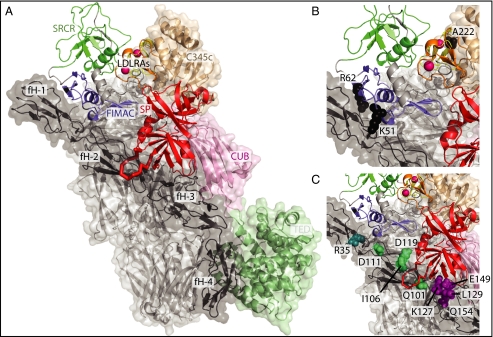
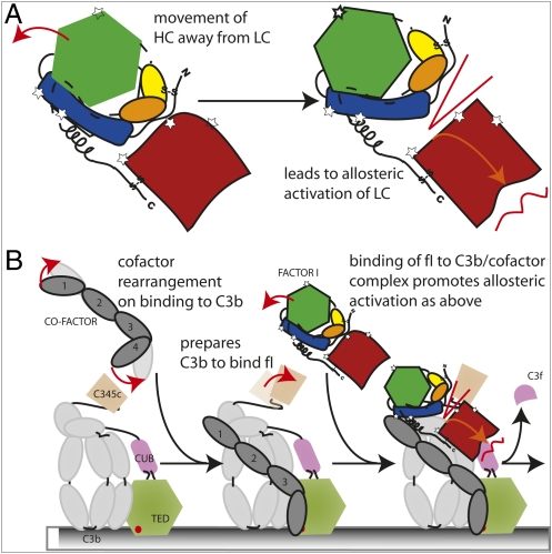
The complement system is a key component of innate and adaptive immune responses. Complement regulation is critical for prevention and control of disease. We have determined the crystal structure of the complement regulatory enzyme human factor I (fI). FI is in a proteolytically inactive form, demonstrating that it circulates in a zymogen-like state despite being fully processed to the mature sequence. Mapping of functional data from mutants of fI onto the structure suggests that this inactive form is maintained by the noncatalytic heavy-chain allosterically modulating activity of the light chain. Once the ternary complex of fI, a cofactor and a substrate is formed, the allosteric inhibition is released, and fI is oriented for cleavage. In addition to explaining how circulating fI is limited to cleaving only C3b/C4b, our model explains the molecular basis of disease-associated polymorphisms in fI and its cofactors.


Conflict of interest statement

The authors declare no conflict of interest.

Figures

Fig. 1.
Fig. 1.
The structure of human fI. (A) Cartoon schematic of fI. FIMAC is shown in blue, SRCR in green, LDLRA1 in yellow, LDLRA2 in orange, and SP in red; unmodeled loops are shown by dashed lines; N-linked glycosylation sites are shown by white stars. (B and C) The protein is shown in two views as a cartoon representation with a transparent surface. Disulfide bonds and two bound Ca2+ ions are shown as ball-and-stick representations. The six glycosylated Asn residues and attached GlcNAc residues (cyan) are shown as stick representations. Domains are colored as A.
Fig. 2.
Fig. 2.
Structural details of human fI. (A) The four crystallographically independent copies of fI are overlaid in cartoon representations revealing no major variation in packing between the heavy and light chains. B and C show the density (weighted 2FO-FC) for the bound Ca2+ in LDLRA2 and novel disulphide between residues 15 and 237 in copy A.
Fig. 3.
Fig. 3.
Zymogenicity of the fI serine protease domain. Overlay of trypsin (Protein Data Bank ID 3MI4) (green and purple) with the fI serine protease domain (gray). The catalytic triad of trypsin is shown as red sticks, and the region of the active site is circled in red. The activation domain loops and N terminus, which are mobile and disordered in trypsinogen (25), are shown in purple and are labeled. Surrounding the overlay, the Cα trace of each component of the trypsin activation domain (purple: activation domain; green: surrounding region) is overlaid with the coordinates of the equivalent regions from the four independent molecules of fI. The fI N terminus is disordered in all but one copy of the molecule, and even here it is mobile (average B factor, 85 Å2) and is not in an active conformation. Factor I activation loops 1 and 2 are not visible in the electron density in any of the four copies, and activation loop 3 can be built only in two copies (average B factor, 66 Å2).
Fig. 4.
Fig. 4.
Mapping of heavy-chain mutations that alter fI activity. (A) Residues corresponding to mutations that impair secretion or activity. The fI heavy chain is shown as in Fig. 1C. Mutations that disrupt the fold of the heavy chain [cyan (21, 22)], or impair the fI catalytic activity [teal (21, 33)] are shown in ball representation. The V134M mutant (32), for which no available secretion or activity data are available, also is shown in green. The arrow points to the face of the heavy chain in contact with the light chain. (B) Mutations that increase the rate of activity (21, 22) of the enzyme cluster around the N terminus of the heavy chain and its region of contact with the light chain. The fI heavy chain is shown as a gray surface, except for its N terminus, which is represented by a gray ribbon. The black line marks the footprint of the light chain onto the heavy chain.
Fig. 5.
Fig. 5.
Modeling the ternary complex of C3b, fH1–4, and fI. Model for the C3b:fH1–4:fI ternary complex in the arrangement leading to the first C3b cleavage. (A) Model of the C3b:fH1–4:fI ternary complex. FI is shown as a cartoon; C3b and fH1–4 are shown as cartoons and semitransparent surfaces The C3b representation is adapted from ref. : C345C domain is shown in light bronze; the CUB domain in pink, the thioester containing domain in light green; and the linker domain, C3 α′ N-terminal domain, and macroglobulin domains of C3b in light gray. (B) Residues corresponding to surface mutations on fI that impair activity and contact C3b (36, 37) or cofactor (21) in the model for the ternary complex. (C) Zoom view of the contact of fI with the cofactor complement control protein domain2–3 junction. The mobile loop in fI (residues 419–425, main chain shown in red) constitutes a point of contact with cofactor surface residues that either impair cofactor activity [vaccine complement control protein (11, 42) shown in green; C4bp (49) shown in purple] or are sites of disease-associated mutations [fH (7, 41) shown in cyan].
Fig. 6.
Fig. 6.
Schematic of the proposed cofactor model. Red arrows indicate domain rearrangements. (A) Cartoon of the proposed allosteric activation of the SP domain via alteration of the heavy-chain (HC)/light-chain (LC) interface. (B) Schematic of the assembly of the C3b:fH1–4:fI ternary complex, colored as in Fig. 5, extended from cartoon of cofactor–C3b interactions (43).

References

    1. Ricklin D, Hajishengallis G, Yang K, Lambris JD. Complement: A key system for immune surveillance and homeostasis. Nat Immunol. 2010;11:785–797. - PMC - PubMed
    1. Lachmann PJ, Müller-Eberhard HJ. The demonstration in human serum of “conglutinogen-activating factor” and its effect on the third component of complement. J Immunol. 1968;100:691–698. - PubMed
    1. Harris CL, Morgan BP. Complement Regulatory Proteins. London: Academic; 1999.
    1. Nicol PA, Lachmann PJ. The alternate pathway of complement activation. The role of C3 and its inactivator (KAF) Immunology. 1973;24:259–275. - PMC - PubMed
    1. van Lookeren Campagne M, Wiesmann C, Brown EJ. Macrophage complement receptors and pathogen clearance. Cell Microbiol. 2007;9:2095–2102. - PubMed

Publication types

MeSH terms

Associated data

LinkOut - more resources