. 2011 Aug 2;108(31):12764-9.
doi: 10.1073/pnas.1015013108. Epub 2011 Jul 18.

Genetic analysis of specific and redundant roles for p38alpha and p38beta MAPKs during mouse development

Affiliations

Genetic analysis of specific and redundant roles for p38alpha and p38beta MAPKs during mouse development

Ivan del Barco Barrantes et al. Proc Natl Acad Sci U S A. .

Abstract

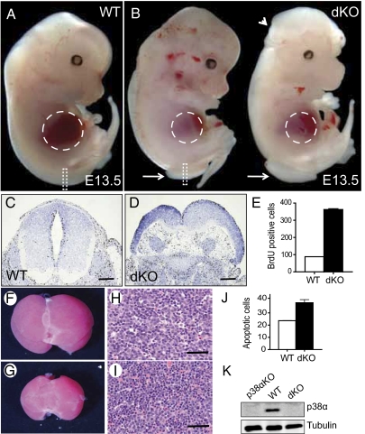
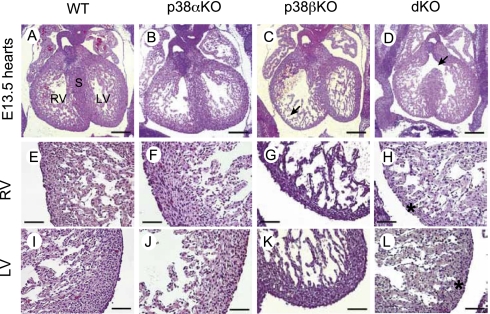
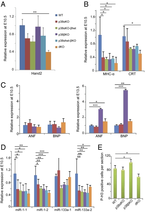
p38α MAPK is an important regulator of cellular responses induced by external cues, but the elucidation of physiological functions for p38α has been complicated by the possible functional redundancy in vivo with the related family member p38β. We found that mice with combined deletion of p38α and p38β display diverse developmental defects at midgestation, including major cardiovascular abnormalities, which are observed neither in single knockout nor in double heterozygous embryos. Expression analysis indicates specific functions of p38α and p38β in the regulation of cardiac gene expression during development. By using knock-in animals that express p38β under control of the endogenous p38α promoter, we also found that p38β cannot perform all of the functions of p38α during embryogenesis. Our results identify essential roles for p38α and p38β during development and suggest that some specific functions may be explained by differences in expression patterns.


Conflict of interest statement

The authors declare no conflict of interest.

Figures

Fig. 1.
Fig. 1.
Developmental defects in p38α and p38β knockout embryos. (A and B) Wild-type (WT) and p38α/Δ)p38β(−/−) Sox2-Cre (dKO) E13.5 embryos are shown. Dashed lines outline the liver, arrows indicate spina bifida, and the arrowhead denotes exencephaly. (C and D) BrdU staining of the neural tube sections corresponding to the areas outlined by dashed rectangles in A and B. (E) Quantification of the BrdU staining in neural tubes (three sections of each genotype). (Scale bars: 500 μm.) (F and G) Liver gross morphology. (H and I) Haematoxylin and eosin staining in liver sections. (Scale bars: 50 μm.) (J) Quantification of TUNEL-positive cells in livers (three sections of each genotype). (K) Deletion of p38α by Sox2-Cre was analyzed by immunoblotting in whole lysates of E13.5 embryos. Error bars indicate SD.
Fig. 2.
Fig. 2.
Cardiac defects in p38α and p38β knockout embryos. (AD) Hematoxylin and eosin staining of transverse sections of hearts from wild-type (WT), p38α/Δ) Sox2-Cre (p38αKO), p38β(−/−) (p38βKO), and p38α/Δ)p38β(−/−) Sox2-Cre (dKO) E13.5 littermate embryos. Note that ventricular septal defects are observed in double KO hearts (D, arrow). (EH) Higher magnification of right ventricles showing thinner compact myocardium in p38β KO and dKO hearts (asterisk). (IL) Higher magnification of left ventricles showing thinner myocardium in dKO hearts (asterisk). S, septum; RV, right ventricle; LV, right ventricle. (Scale bars: AD, 500 μm; EL, 100 μm.)
Fig. 3.
Fig. 3.
Modulation of cardiac gene expression and proliferation. Expression of the indicated genes was analyzed by quantitative RT-PCR in biological triplicates of the following genotypes: wild-type (WT), p38α/Δ) Sox2-Cre (p38αKO), p38α/Δ) p38β(−/−) Sox2-Cre (p38αKO-βhet), p38β(−/−) (p38βKO), p38α/+) p38β(−/−) Sox2-Cre (p38αhet-βKO), and p38α/Δ)p38β(−/−) Sox2-Cre (dKO). RNA was isolated from three individual hearts in E10.5 embryos, and from three pools of three hearts each (nine hearts total) in E13.5 embryos. (A) Hand2 transcription factor is down-regulated in E10.5 dKO hearts. (B) Contractile protein MHC-α is down-regulated in all mutant hearts, whereas the differentiation marker CRT is down-regulated in dKO hearts. (C) Up-regulation of the hypertrophic markers ANF and BNP in p38βKO and dKO hearts at E13.5 but not at E10.5. (D) Down-regulation of miR-1-1, miR-1-2, and miR-133a-2 in all mutant hearts. (E) Phospho-histone H3 (P-H3) staining of E13.5 heart sections. MHC-α, myosin heavy chain-α; CRT; calreticulin; ANF, atrial natrium factor; BNP, B-type natriuretic protein. Error bars indicate SD. Statistical significance (n = 3) was determined by using one-way ANOVA-Tukey's test. Changes are referred to the expression levels in WT hearts (given the value of 1 for each gene) and indicated as *P < 0.05, **P < 0.01, and ***P < 0.001.
Fig. 4.
Fig. 4.
Generation of mice expressing p38β under control of the endogenous p38α promoter (p38βKIα allele). (A) RT-PCR analysis of p38α mRNA extracted from the indicated WT adult tissues. (B) RT-PCR analysis of p38β mRNA extracted from the indicated adult tissues of mice wild-type (WT), p38β(−/−) (p38βKO), and p38αKI/+)p38β(−/−) (KIp38βKO).
Fig. 5.
Fig. 5.
Expression of p38β under control of the endogenous p38α promoter does not rescue cardiac defects in embryos. (A) Immunoblotting of p38β protein in the indicated whole embryos and embryo tissues wild-type (WT), p38α/Δ)Sox2-Cre (p38αKO), p38β(−/−) (p38βKO), and p38αKI/+)p38β(−/−) (KIp38βKO, one copy p38α, and the other p38βKIα). (B) Quantitative RT-PCR analysis of p38β mRNA expression in heart, liver, and lung of E13.5 embryos p38αKO, p38βKO, and p38αKI/Δ p38βKO, which express p38β only under control of the p38α promoter (p38αKI/Δ)p38β(−/−)Sox2-Cre). Expression levels of p38β were referred to those in WT tissues, which were given the value of 1. (C) Histological analysis showing that p38αKI/Δ p38βKO embryos have similar cardiac defects as the p38α and p38β double knockout embryos.

References

    1. Cuadrado A, Nebreda AR. Mechanisms and functions of p38 MAPK signalling. Biochem J. 2010;429:403–417. - PubMed
    1. Adams RH, et al. Essential role of p38alpha MAP kinase in placental but not embryonic cardiovascular development. Mol Cell. 2000;6:109–116. - PubMed
    1. Mudgett JS, et al. Essential role for p38alpha mitogen-activated protein kinase in placental angiogenesis. Proc Natl Acad Sci USA. 2000;97:10454–10459. - PMC - PubMed
    1. Hui L, et al. p38alpha suppresses normal and cancer cell proliferation by antagonizing the JNK-c-Jun pathway. Nat Genet. 2007;39:741–749. - PubMed
    1. Beardmore VA, et al. Generation and characterization of p38beta (MAPK11) gene-targeted mice. Mol Cell Biol. 2005;25:10454–10464. - PMC - PubMed

Publication types

MeSH terms

Substances