. 2011 Aug 2;108(31):12752-7.
doi: 10.1073/pnas.1006437108. Epub 2011 Jul 18.

Deletion of mouse Porcn blocks Wnt ligand secretion and reveals an ectodermal etiology of human focal dermal hypoplasia/Goltz syndrome

Affiliations

Deletion of mouse Porcn blocks Wnt ligand secretion and reveals an ectodermal etiology of human focal dermal hypoplasia/Goltz syndrome

Jared J Barrott et al. Proc Natl Acad Sci U S A. .

Abstract

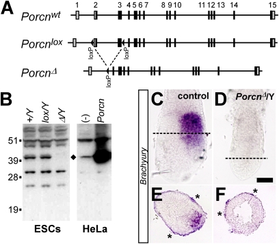
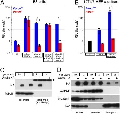
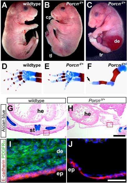
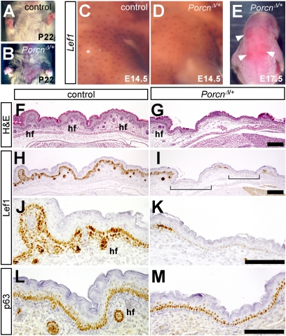
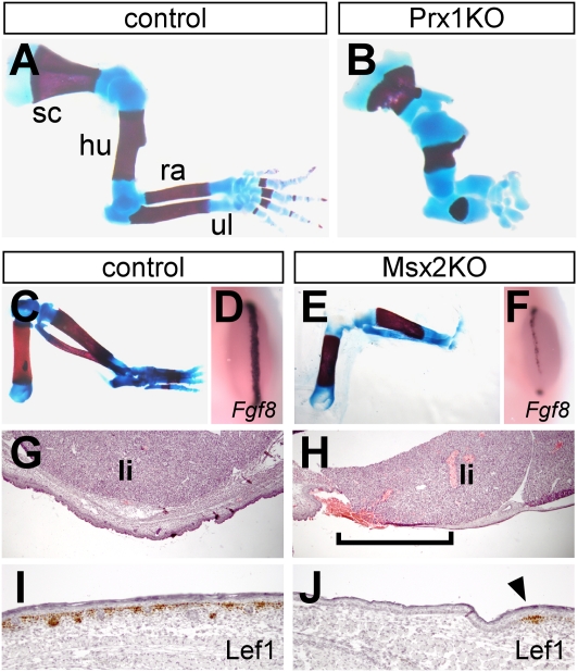
The Drosophila porcupine gene is required for secretion of wingless and other Wnt proteins, and sporadic mutations in its unique human ortholog, PORCN, cause a pleiotropic X-linked dominant disorder, focal dermal hypoplasia (FDH, also known as Goltz syndrome). We generated a conditional allele of the X-linked mouse Porcn gene and analyzed its requirement in Wnt signaling and embryonic development. We find that Porcn-deficient cells exhibit a cell-autonomous defect in Wnt ligand secretion but remain responsive to exogenous Wnts. Consistent with the female-specific inheritance pattern of FDH, Porcn hemizygous male embryos arrest during early embryogenesis and fail to generate mesoderm, a phenotype previously associated with loss of Wnt activity. Heterozygous Porcn mutant females exhibit a spectrum of limb, skin, and body patterning abnormalities resembling those observed in human patients with FDH. Many of these defects are recapitulated by ectoderm-specific deletion of Porcn, substantiating a long-standing hypothesis regarding the etiology of human FDH and extending previous studies that have focused on downstream elements of Wnt signaling, such as β-catenin. Conditional deletion of Porcn thus provides an experimental model of FDH, as well as a valuable tool to probe Wnt ligand function in vivo.


Conflict of interest statement

The authors declare no conflict of interest.

Figures

Fig. 1.
Fig. 1.
Generating and characterizing a conditional mouse Porcn allele. (A) Schematic diagram of WT, loxP-targeted, and deletion alleles of Porcn. Exons are boxed and numbered, with the coding region indicated in black. (B) Western blot of whole-cell lysates from parental and targeted ES cells (Left) and HeLa cells transfected with empty vector or mouse Porcn (Right) using a polyclonal antiserum against a C-terminal Porcn epitope. The diamond indicates the band corresponding to overexpressed mouse Porcn and is present only in parental and Porcnlox ES cells. (C and D) Whole-mount in situ hybridization for Brachyury (purple) on E6.5 control or PorcnΔ/Y embryos. (Scale bar: 100 μm.) (E and F) Sections through Brachyury whole-mount stained embryos, with approximate positions indicated by the dotted lines in C and D. Note that sections were taken from an independent representative pair of embryos, overstained to preserve signal in sections (asterisks indicate nonspecific background). PorcnΔ/Y embryos consistently contain a hollow lumen at this stage, indicating a lack of gastrulation.
Fig. 2.
Fig. 2.
Wnt activity and processing in Porcn-deficient cells. (A) TOPFlash luciferase reporter assays comparing the response of Porcnlox/Y (blue) and PorcnΔ/Y (red) ES cells with exogenous Wnt3a (10% (vol/vol) l-Wnt3a–conditioned medium) or endogenously overexpressed Wnt3a (cells transfected with Wnt3a expression plasmid). Where indicated, cells were cotransfected with expression plasmids for WT or H341L mutant human PORCN. Relative light units indicate TOPFlash activity, normalized to an internal transfection control and plotted on a log scale as fold change relative to untreated Porcnlox/Y cells (n = 3–8 independent experiments per condition). RLU, relative light unit. *P < 0.05 by Welch's two-tailed t test. (B) Firefly luciferase assays of 10T1/2 cells stably infected with a TOPFlash-based lentiviral reporter, cocultured with immortalized Porcnlox/Y (blue) and PorcnΔ/Y (red) MEFs that were previously infected with an empty retroviral vector (LNCX) or LNC-Wnt3a-HA. Relative light units are plotted as fold change relative to reporter-transduced cells cultured alone (black) (n = 3 independent experiments). *P < 0.05 by Welch's two-tailed t test. (C) Western blots (with antibodies indicated to left of panels) on whole-cell lysates from control and Wnt3a-HA–expressing MEFs (Left) and on anti-HA immunoprecipitates of conditioned media from the same cells (Right, asterisk indicates rabbit IgG heavy chain). Note that the immunoprecipitates represent ∼10-fold more input material than the corresponding cellular lysates. Tubulin serves as a loading control. (D) Western blots (antibodies indicated on left) on lysates from control and Wnt3a-HA–expressing MEFs, either unprocessed (whole) or separated into aqueous and detergent phases by Triton X-114 extraction. GAPDH and β-catenin serve as controls for recovery of nonacylated aqueous-phase proteins, and Ras serves as a control for detergent-phase recovery of acylated proteins.
Fig. 3.
Fig. 3.
FDH-like phenotypes in PorcnΔ/+ heterozygous embryos. (AC) Comparing E17.5 PorcnΔ/+ embryos with WT reveals a range of phenotypes that include cleft palate, tail hypoplasia, omphalocele, atrophic dermis through which the liver is visible, and tail/posterior axis truncation. cp, cleft palate; de, atrophic dermis; om, omphalocele; tr, tail/posterior axis truncation; tl, tail hypoplasia. (DF) Alcian blue/alizarin red skeletal stains of WT and PorcnΔ/+ forelimbs. The arrow in F indicates lack of autopod, accompanied by absence of ulna. (GH) Alcian blue/nuclear fast red staining to reveal skeletal elements, including sternum, of E15.5 WT and PorcnΔ/+ ventral body walls sectioned at the level of the heart. he, heart; st, sternum. (I and J) Immunostaining of sections semiadjacent to G and H (approximate positions indicated by red boxes) for E-cadherin (red) and the dermis/mesenchyme marker PDGF receptor-α (green). de, dermis; ep, epidermis. (Scale bars: G and H, 500 μm; I and J, 50 μm.)
Fig. 4.
Fig. 4.
Focal absence of hair follicles in PorcnΔ/+ heterozygotes. (A and B) Ventral view of WT and PorcnΔ/+ littermates at weanling stage (postnatal day 22). (CE) Whole-mount in situ hybridization for Lef1 (brown) on shoulder region of E14.5 control or PorcnΔ/+ embryos. The asterisk indicates a fragment of skin accidentally removed during dissection. (E) Dorsal view of an E17.5 PorcnΔ/+ embryo, indicating patches of unusually smooth skin (arrowheads). (FM) Semiadjacent sections of dorsal skin from E17.5 WT or PorcnΔ/+ embryos, stained with H&E or immunostained for Lef1 or p63. The brackets in I indicate patches devoid of hair follicles and expressing low or no Lef1. hf, hair follicle. (Scale bars: 100 μm.)
Fig. 5.
Fig. 5.
Tissue-specific Porcn deletion phenotypes. (A and B) Alcian blue/alizarin red-stained forelimbs of E17.5 control or Porcnlox/Y; Prx1-Cre (Prx1KO) embryos. Mutant embryos exhibit shortening of all skeletal elements and loss of distal digits. hu, humerus; ra, radius; sc, scapula; ul, ulna. (CF) Hind-limb skeleton preparations of E18.5 control or Porcnlox/Y; Msx2-Cre (Msx2KO) embryos and whole-mount in situ hybridization for the AER marker Fgf8 on E11.5 hind limbs. Note the almost complete absence of autopod in E18.5 mutant and focal loss of Fgf8 expression at E11.5. (G and H) H&E-stained sections of the ventral body wall from E18.5 control or Msx2KO embryos. The brackets in H indicate an area of severe dermal thinning in mutant body wall, such that liver almost directly abuts surface ectoderm. li, liver. (I and J) Lef1 immunostaining of dorsal skin from E18.5 control or Msx2KO embryos, revealing extensive domain of hairless Lef1-devoid epidermis in mutant (arrowhead in J indicates isolated patch of Lef1+ basal cells). (Scale bars: G and H, 500 μm; I and J, 100 μm.)

References

    1. Logan CY, Nusse R. The Wnt signaling pathway in development and disease. Annu Rev Cell Dev Biol. 2004;20:781–810. - PubMed
    1. MacDonald BT, Tamai K, He X. Wnt/beta-catenin signaling: Components, mechanisms, and diseases. Dev Cell. 2009;17:9–26. - PMC - PubMed
    1. Grigoryan T, Wend P, Klaus A, Birchmeier W. Deciphering the function of canonical Wnt signals in development and disease: Conditional loss- and gain-of-function mutations of beta-catenin in mice. Genes Dev. 2008;22:2308–2341. - PMC - PubMed
    1. Veeman MT, Axelrod JD, Moon RT. A second canon. Functions and mechanisms of beta-catenin-independent Wnt signaling. Dev Cell. 2003;5:367–377. - PubMed
    1. Nelson WJ, Nusse R. Convergence of Wnt, beta-catenin, and cadherin pathways. Science. 2004;303:1483–1487. - PMC - PubMed

Publication types

MeSH terms