. 2011 Aug 2;108(31):12805-10.
doi: 10.1073/pnas.1102298108. Epub 2011 Jul 18.

SPR-5 is a histone H3K4 demethylase with a role in meiotic double-strand break repair

Affiliations

SPR-5 is a histone H3K4 demethylase with a role in meiotic double-strand break repair

Amanda C Nottke et al. Proc Natl Acad Sci U S A. .

Abstract

Regulation of histone methylation levels has long been implicated in multiple cellular processes, many of which involve transcription. Here, however, we report a unique role for the Caenorhabditis elegans histone demethylase SPR-5 in meiotic DNA double-strand break repair (DSBR). SPR-5 shows enzymatic activity toward H3K4me2 both in vitro and in the nematode germline, and spr-5 mutants show several phenotypes indicating a perturbation of DSBR, including increased p53-dependent germ cell apoptosis, increased levels of the DSBR marker RAD-51, and sensitivity toward DSB-inducing treatments. spr-5 mutants show no transcriptional misregulation of known DSBR involved genes. Instead, SPR-5 shows a rapid subcellular relocalization upon DSB-inducing treatment, which suggests that SPR-5 may function directly in DSBR.


Conflict of interest statement

The authors declare no conflict of interest.

Figures

Fig. 1.
Fig. 1.
SPR-5 is an H3K4me2 demethylase that functions in the C. elegans germline. (A and B) SPR-5 demethylates H3K4me2. Full-length FLAG-SPR-5 was incubated with purified calf thymus histones (A) or whole-worm protein lysate (B). Western blots were used to separate samples, then probed using anti-H3K4me2 antibodies and Ponceau dye (Upper) or anti-tubulin antibody (Lower) for loading controls. (C) Schematic of spr-5 mutant alleles. spr-5(by134) contains an early nonsense mutation within the TOWER domain (light gray). spr-5(by101) contains a transposon insertion (Tc3) adjacent to predicted catalytic residues in the amine oxidase domain (medium gray). The SWIRM domain (dark gray), predicted for protein–protein interactions, is unaffected. (D) spr-5 mutants show increased germline H3K4me2. Gonads were dissected from 50 animals per genotype. After separation by Western blot, samples were probed with anti-H3K4me2 and anti-tubulin (loading control) antibodies. (E) Immunostaining reveals that spr-5 mutant germ cells show increased H3K4me2. Gonads from either GFP::H2B-expressing animals (control) or spr-5 (F70+) worms were immunostained for H3K4me2 (Left and Center). (Scale bars, 2 μm.) Individual nuclei were sampled for fluorescent intensity using ImageJ (Right). The bars represent the mean fluorescent intensity. (F) Germ cell H3K4me2 is progressively misregulated in spr-5 mutants. Germline H3K4me2 immunostaining was compared between wild-type and both late-generation (F70+) and early-generation (F3, created from a single outcross of the F70+ strain) spr-5 mutants. (Scale bars, 5 μm.)
Fig. 2.
Fig. 2.
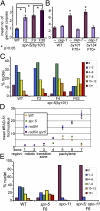
spr-5 mutants show a progressive defect in DNA double-strand break repair. (A) spr-5 mutants show a progressive increase in germ cell apoptosis. Error bars represent the SEM. P values were determined using the two-tailed Mann-Whitney test, 95% confidence interval. (B) Increased apoptosis in late generation spr-5 mutants is cep-1-dependent. Wild-type as well as late-generation spr-5(by101) and spr-5(by134) animals were examined after growth in either control(RNAi) or cep-1(RNAi) plates. As reported previously, RNAi treatment (both control and target gene-specific) results in higher levels of germ cell apoptosis (approximately three apoptotic germ cells in the control) (31), but the relative increase in spr-5 mutants remains. Error bars represent the SEM; P values were determined by the two-tailed Mann-Whitney test, 95% confidence interval. (C) spr-5 mutants show a progressive increase in mid-pachytene RAD-51 foci. The number of RAD-51 foci per nucleus was quantified in the mid-pachytene zone (zone 5 as in ref. and 20) for at least three gonads per strain. Ranges (number of foci detected per nucleus) and color codes are indicated in the key. The rightward shift of the histograms indicates a higher percentage of nuclei with more RAD-51 foci. (D) Analysis of repair-defective mutants of the indicated genotypes reveals no difference in the number of DSBs formed. The total number of RAD-51 foci in each nucleus was quantified as in C and Fig. S3. However, here the data are displayed as the mean number of RAD-51 foci per nucleus (y axis) in each zone or region of the germline (x axis) for at least three gonads per genotype. There is no statistically significant difference between rad-54 and rad-54 spr-5 mutants at any stage. Experiments were done on generation F3–F5 mutants. (E) The increase in RAD-51 foci is dependent on the meiotic endonuclease spo-11. Histograms depict the quantitation of RAD-51 foci in midpachytene (zone 5) nuclei for at least three germlines for each indicated genotype.
Fig. 3.
Fig. 3.
SPR-5 relocalizes in response to DNA double-strand breaks and is required for IR resistance. (A) SPR-5 relocalizes rapidly upon irradiation. Gonads were dissected from 24 h post-L4 wild-type worms 15–30 min after exposure to 60 Gy of IR and compared with untreated controls (no tx). Images are from pachytene nuclei. (Scale bars, 5 um.) (B) SPR-5 relocalizes upon DSB-inducing IR but not in response to other DNA damage-inducing reagents. Animals were treated as in A with 60 Gy for the IR, with 200 J/m2 for the UV and 500 nM for the CPT treatments. (C) Early-generation (F2) spr-5(by101) mutants show decreased embryonic viability compared with wild-type after treatment with the indicated doses of IR, but not UV-C or CPT. At least 30 animals were assayed per genotype and condition. Points represent the mean embryonic survival, error bars represent the SEM.
Fig. 4.
Fig. 4.
spr-5 germline gene targets are under-enriched for germline-expressed genes and do not include known DSBR genes. (A) Comparison of genes up-regulated in spr-5 mutants with published germline function and expression data (32). Germline expression refers to genes enriched in wild-type hermaphrodites compared with germline-deficient glp-4 mutants. Somatic expression refers to genes of each functional class that are not germline-enriched. Genes important for either oogenesis (Oo.) or spermatogenesis function (Sp.) were determined by comparing oocyte-only producing mutants [fem-1(lf)] to sperm-only producing mutants [fem-3(gf)] or vice versa (32). Other genes with germline expression are those genes that are germline-enriched but with neither oogenesis nor spermatogenesis known functions. Enrichment scores are statistically significant (P < 0.05). (B) Comparison of spr-5 gene targets with other published datasets. Four tissue-specific datasets showed statistically significant overrepresentation of spr-5 gene targets within the datasets. i. (33); ii. (34); iii. (35); iv. (36). Enrichment scores are statistically significant (P < 0.05). (C) Examination of DSBR repair gene expression levels in spr-5 mutants. qRT-PCR was performed on samples confirmed in Fig. S5 for candidate DSBR genes.
Fig. 5.
Fig. 5.
Model for DSBR progression defect phenotype in both early- and late-generation spr-5 mutants. In wild-type animals, we propose that SPR-5 acts after DSBs occur to remove H3K4me2 (green flags), allowing DSBR factors to complete repair efficiently (Left). In early-generation spr-5 mutants, the global levels of H3K4me2 are minimally affected, but after DSBs occur H3K4me2 is not removed by SPR-5, leading to inefficient DSBR (Center). In late-generation spr-5 mutants, the lack of SPR-5 to act after damage and the general increase in H3K4me2 combine to further impair DSBR progression (Right).

Similar articles

Cited by

References

    1. Martin C, Zhang Y. The diverse functions of histone lysine methylation. Nat Rev Mol Cell Biol. 2005;6:838–849. - PubMed
    1. Ng SS, Yue WW, Oppermann U, Klose RJ. Dynamic protein methylation in chromatin biology. Cell Mol Life Sci. 2009;66:407–422. - PMC - PubMed
    1. Myers S, et al. Drive against hotspot motifs in primates implicates the PRDM9 gene in meiotic recombination. Science. 2010;327:876–879. - PMC - PubMed
    1. Buard J, Barthès P, Grey C, de Massy B. Distinct histone modifications define initiation and repair of meiotic recombination in the mouse. EMBO J. 2009;28:2616–2624. - PMC - PubMed
    1. Baudat F, et al. PRDM9 is a major determinant of meiotic recombination hotspots in humans and mice. Science. 2010;327:836–840. - PMC - PubMed

Publication types

MeSH terms