

Sec16B is involved in the endoplasmic reticulum export of the peroxisomal membrane biogenesis factor peroxin 16 (Pex16) in mammalian cells
- PMID: 21768384
- PMCID: PMC3150892
- DOI: 10.1073/pnas.1103283108
Sec16B is involved in the endoplasmic reticulum export of the peroxisomal membrane biogenesis factor peroxin 16 (Pex16) in mammalian cells
Abstract
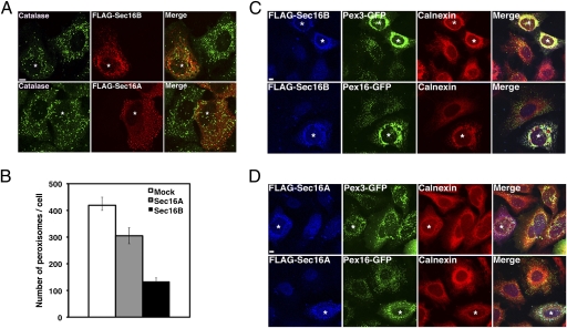
Sec16 plays a key role in the formation of coat protein II vesicles, which mediate protein transport from the endoplasmic reticulum (ER) to the Golgi apparatus. Mammals have two Sec16 isoforms: Sec16A, which is a longer primary ortholog of yeast Sec16, and Sec16B, which is a shorter distant ortholog. Previous studies have shown that Sec16B, as well as Sec16A, defines ER exit sites, where coat protein II vesicles are formed in mammalian cells. Here, we reveal an unexpected role of Sec16B in the biogenesis of mammalian peroxisomes. When overexpressed, Sec16B was targeted to the entire ER, whereas Sec16A was mostly cytosolic. Concomitant with the overexpression of Sec16B, peroxisomal membrane biogenesis factors peroxin 3 (Pex3) and Pex16 were redistributed from peroxisomes to Sec16B-positive ER membranes. Knockdown of Sec16B but not Sec16A by RNAi affected the morphology of peroxisomes, inhibited the transport of Pex16 from the ER to peroxisomes, and suppressed expression of Pex3. These phenotypes were significantly reversed by the expression of RNAi-resistant Sec16B. Together, our results support the view that peroxisomes are formed, at least partly, from the ER and identify a factor responsible for this process.
Conflict of interest statement
The authors declare no conflict of interest.
Figures


Similar articles
-
Dual function of Sec16B: Endoplasmic reticulum-derived protein secretion and peroxisome biogenesis in mammalian cells.Cell Logist. 2011 Jul;1(4):164-167. doi: 10.4161/cl.1.4.18341. Epub 2011 Jul 1. Cell Logist. 2011. PMID: 22279616 Free PMC article.
-
Characterization of human Sec16B: indications of specialized, non-redundant functions.Sci Rep. 2011;1:77. doi: 10.1038/srep00077. Epub 2011 Aug 30. Sci Rep. 2011. PMID: 22355596 Free PMC article.
-
A New Yeast Peroxin, Pex36, a Functional Homolog of Mammalian PEX16, Functions in the ER-to-Peroxisome Traffic of Peroxisomal Membrane Proteins.J Mol Biol. 2017 Nov 24;429(23):3743-3762. doi: 10.1016/j.jmb.2017.10.009. Epub 2017 Oct 14. J Mol Biol. 2017. PMID: 29037759 Free PMC article.
-
De novo peroxisome biogenesis: Evolving concepts and conundrums.Biochim Biophys Acta. 2016 May;1863(5):892-901. doi: 10.1016/j.bbamcr.2015.09.014. Epub 2015 Sep 14. Biochim Biophys Acta. 2016. PMID: 26381541 Free PMC article. Review.
-
Targeting and insertion of peroxisomal membrane proteins: ER trafficking versus direct delivery to peroxisomes.Biochim Biophys Acta. 2016 May;1863(5):870-80. doi: 10.1016/j.bbamcr.2015.09.021. Epub 2015 Sep 25. Biochim Biophys Acta. 2016. PMID: 26392202 Review.
Cited by
-
Bi-allelic mutation in SEC16B alters collagen trafficking and increases ER stress.EMBO Mol Med. 2023 Apr 11;15(4):e16834. doi: 10.15252/emmm.202216834. Epub 2023 Mar 14. EMBO Mol Med. 2023. PMID: 36916446 Free PMC article.
-
MT1-MMP recruits the ER-Golgi SNARE Bet1 for efficient MT1-MMP transport to the plasma membrane.J Cell Biol. 2019 Oct 7;218(10):3355-3371. doi: 10.1083/jcb.201808149. Epub 2019 Sep 13. J Cell Biol. 2019. PMID: 31519727 Free PMC article.
-
PEX16: a multifaceted regulator of peroxisome biogenesis.Front Physiol. 2013 Sep 3;4:241. doi: 10.3389/fphys.2013.00241. Front Physiol. 2013. PMID: 24027535 Free PMC article. Review.
-
A new pathway for mitochondrial quality control: mitochondrial-derived vesicles.EMBO J. 2014 Oct 1;33(19):2142-56. doi: 10.15252/embj.201488104. Epub 2014 Aug 8. EMBO J. 2014. PMID: 25107473 Free PMC article. Review.
-
Novel genomic variants related to visceral adiposity index (VAI) and body adiposity index (BAI) in Indian sib-pairs.Int J Obes (Lond). 2024 Nov;48(11):1552-1558. doi: 10.1038/s41366-024-01570-y. Epub 2024 Jul 7. Int J Obes (Lond). 2024. PMID: 38971891
References
-
- van den Bosch H, Schutgens RB, Wanders RJ, Tager JM. Biochemistry of peroxisomes. Annu Rev Biochem. 1992;61:157–197. - PubMed
-
- Fagarasanu A, Fagarasanu M, Rachubinski RA. Maintaining peroxisome populations: A story of division and inheritance. Annu Rev Cell Dev Biol. 2007;23:321–344. - PubMed
-
- Lazarow PB, Fujiki Y. Biogenesis of peroxisomes. Annu Rev Cell Biol. 1985;1:489–530. - PubMed
-
- Fujiki Y, Okumoto K, Kinoshita N, Ghaedi K. Lessons from peroxisome-deficient Chinese hamster ovary (CHO) cell mutants. Biochim Biophys Acta. 2006;1763:1374–1381. - PubMed
Publication types
MeSH terms
Substances
LinkOut - more resources
Full Text Sources
Molecular Biology Databases
