. 2011 Aug 2;108(31):12863-8.
doi: 10.1073/pnas.1105002108. Epub 2011 Jul 18.

High-resolution, noninvasive longitudinal live imaging of immune responses

Affiliations

High-resolution, noninvasive longitudinal live imaging of immune responses

Midhat H Abdulreda et al. Proc Natl Acad Sci U S A. .

Abstract

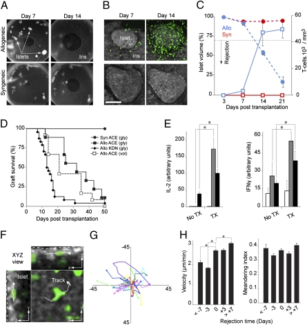
Intravital imaging emerged as an indispensible tool in biological research, and a variety of imaging techniques have been developed to noninvasively monitor tissues in vivo. However, most of the current techniques lack the resolution to study events at the single-cell level. Although intravital multiphoton microscopy has addressed this limitation, the need for repeated noninvasive access to the same tissue in longitudinal in vivo studies remains largely unmet. We now report on a previously unexplored approach to study immune responses after transplantation of pancreatic islets into the anterior chamber of the mouse eye. This approach enabled (i) longitudinal, noninvasive imaging of transplanted tissues in vivo; (ii) in vivo cytolabeling to assess cellular phenotype and viability in situ; (iii) local intervention by topical application or intraocular injection; and (iv) real-time tracking of infiltrating immune cells in the target tissue.


Conflict of interest statement

Conflict of interest statement: P.-O.B. is one of the founders of the biotech company Biocrine, which is going to use the anterior chamber of the eye as a commercial servicing platform. A.C. and I.L. are also involved in the commercialization of this servicing platform.

Figures

Fig. 1.
Fig. 1.
Transplantation into the anterior chamber (ACE) of the mouse eye enables longitudinal, noninvasive imaging and tracking of immune cells in individual islet grafts in vivo. (A) Images of mouse eyes transplanted with allogeneic or syngeneic islets that have engrafted on the iris. Allogeneic islets disappeared by day 14 after transplantation because of rejection. (B) In vivo confocal z-stacks (shown as maximum projections) of the same intraocular grafts showing progressive infiltration of allogeneic grafts by GFP-labeled (green) T lymphocytes. (Scale bar, 100 μm.) (C) Change in the average islet volume (circles) vs. the number (squares) of graft-infiltrating T-cells (blue, allogeneic; red, syngeneic). Data based on 5 to 31 islets/time point from 5 allogeneic, and 5 to 22 islets/time point from three syngeneic recipients. Results presented as means ± SEM. (D) Survival curves of islet grafts in the ACE or under the kidney capsule (KDN) based on glycemia (gly) or volume (vol) [Syn ACE: n = 4; Allo ACE (gly): n = 7; Allo ACE (vol): n = 12; Allo KDN: n = 24]. (E) IL-2 and IFN-γ production (ELISPOT) in response to alloantigen challenge in cervical/axillary lymph nodes (n = 3 mice) and spleens (n = 3) of nontransplanted (no TX) or transplanted (TX) animals in the ACE. (F) Snapshot (XYZ view) from a 3D time-lapse recording (20 min; POD +21) showing a tracked cell within an islet allograft (outlined with dotted line). (Scale bars, 10 μm.). (G) Two-dimensional flower plot representation of movement trajectories of individual cells tracked (in 3D) within the graft. (H) Dynamic parameters derived from 3D tracking analysis on infiltrating GFP-labeled T cells. Velocity (average speed/path length) and meandering index (displacement rate/velocity; a measure of movement directionality) were measured during 20-min recordings obtained in the same islet allografts at the indicated time points (± 1 d). Data derived from > 2,000 graft-infiltrating cells and pooled from 23 islets in seven mice. Results presented as means ± SEM (n = 3–6 islets per time point). *P < 0.05.
Fig. 2.
Fig. 2.
In situ labeling of islet cells and graft-infiltrating immune cells in the anterior chamber of the eye in vivo. (A) We injected fluorescence-labeled antibodies directly into the anterior chamber of the eye between POD +8 and POD +23 to reveal the phenotypes of graft-infiltrating immune cells; ∼80% of the graft-infiltrating GFP-labeled T-cells were CD8+. (Scale bar, 5 μm.) (B) Ruffled T cells (green) associated with apoptotic islet cells (Annexin V, red; DAPI, blue) during rejection (Movie S4); ∼70% of graft-infiltrating GFP-labeled T cells contacted apoptotic islet cells. *P < 0.05. (Scale bar, 10 μm.) (C) Endocrine islet cells (outlined; Left) and lytic granules (arrowheads) within GFP-labeled ruffled T-cells (green) were in vivo labeled with Lysotracker (red) (Movie S5). Lytic granules within ruffled T-cells faced target islet cells (Center). Immunohistochemistry of fixed intraocular islet allograft showed the presence of granzyme B/perforin (arrowheads; Right) in a graft-infiltrating ruffled T lymphocyte (green) (Fig. S2C). Shown images are representative of a minimum of triplicate experiments. (Scale bars, 5 μm.) (D) In vivo confocal images (maximum projections) of GFP-labeled T cells within subcapsular pancreatic islet grafts after exposure of the kidney at onset of rejection (confirmed by glycemia) at POD +12. Although we were not able to image the islets because of the thick kidney capsule (gray reflection; Left), we were able to recover GFP signal from infiltrating T-cells (green). [Scale bars, 10 μm (Right), 30 μm (Center), and 100 μm (Left).] (E) Close-ups of round, elongated, and ruffled cells with corresponding movement tracks. (Scale bar, 5 μm.) (F) Number of round, elongated, and ruffled cells within the same islet allografts at different time points. Results pooled from 22 islets (n = 3–6 islets/time point in six mice).
Fig. 3.
Fig. 3.
Intraocular transplantation enables longitudinal noninvasive monitoring after local or systemic pharmacological interventions. (A) Fluorescence confocal images (maximum projections) shown in intensity scale to illustrate morphological changes of allograft-infiltrating T cells before and after TAK-779 (50 μM) or CXCL9/CXCL10 (100 nM) acute treatment at the time of rejection (POD +22). (Scale bar, 40 μm.) Our approach revealed noticeable and reversible changes in the cellular morphology and behavior of individual T cells after successive injection of either TAK-779 or CXCL9/CXCL10 into the same eye. (B) We measured significant changes in the velocity and displacement (10 min) of infiltrating T-cells after these treatments. Data derived from 837 (before), 335 (TAK-779), and 340 (CXCL9/CXCL10) GFP-labeled cells, obtained from three islets (three mice) imaged at POD +13, +16, +21, and +22. (C and D) Systemic TAK-779 treatment (250 μg/day; intraperitoneally) between POD +10 and POD +17 resulted in significantly reduced islet graft infiltration by T lymphocytes and reduced overall movement dynamics. Circles represent islet volume and squares the number of intraislet T-cells in TAK-779-treated (filled symbols; n = 15 islets in three mice) or untreated animals (open symbols; n = 22 islets in five mice). Results presented as means ± SEM . (E) Systemic TAK-779 treatment delayed islet rejection (median survival time based on islet volume loss of ≥ 30% = 42 vs. 21 d). *P < 0.05.

References

    1. Martinic MM, von Herrath MG. Real-time imaging of the pancreas during development of diabetes. Immunol Rev. 2008;221:200–213. - PubMed
    1. Prescher A, Mory C, Martin M, Fiedler M, Uhlmann D. Effect of FTY720 treatment on postischemic pancreatic microhemodynamics. Transplant Proc. 2010;42:3984–3985. - PubMed
    1. Leblond F, Davis SC, Valdés PA, Pogue BW. Pre-clinical whole-body fluorescence imaging: Review of instruments, methods and applications. J Photochem Photobiol B. 2010;98(1):77–94. - PMC - PubMed
    1. Toso C, et al. Clinical magnetic resonance imaging of pancreatic islet grafts after iron nanoparticle labeling. Am J Transplant. 2008;8:701–706. - PubMed
    1. Ntziachristos V. Going deeper than microscopy: The optical imaging frontier in biology. Nat Methods. 2010;7:603–614. - PubMed

Publication types

MeSH terms