

Review
doi: 10.1101/cshperspect.a002774.
RNA granules in germ cells
Affiliations
- PMID: 21768607
- PMCID: PMC3225947
- DOI: 10.1101/cshperspect.a002774
Item in Clipboard
Review
RNA granules in germ cells
Cold Spring Harb Perspect Biol.
.
Abstract
"Germ granules" are cytoplasmic, nonmembrane-bound organelles unique to germline. Germ granules share components with the P bodies and stress granules of somatic cells, but also contain proteins and RNAs uniquely required for germ cell development. In this review, we focus on recent advances in our understanding of germ granule assembly, dynamics, and function. One hypothesis is that germ granules operate as hubs for the posttranscriptional control of gene expression, a function at the core of the germ cell differentiation program.
Figures

Electron micrographs of the major germ granule classes. (A) Perinuclear P granule in a meiotic germ cell of C. elegans. Distinct subdomains are visible: the crest (white arrow) and base (arrowhead) overlying a cluster of nuclear pores (black arrows). Scale bar: 500 nm. (Panel A is adapted from Sheth et al. [2010] and reproduced, with permission, from The Company of Biologists © 2010.) (B) Balbiani body in a mouse oocyte. Black arrow points to mitochondria clustered around Golgi membranes (BB). N, nucleus. Scale bar: 5 um. (Panel B is adapted from Pepling et al. [2007] and reproduced, with permission, from National Academy of Sciences, USA © 2007.) (C) Sponge bodies in the Drosophila nurse cell. Sponge bodies (asterisks) intermingled with mitochondria (m) and ER cisternae (arrows) near an intercellular bridge (black arrowheads) connecting the nurse cell (nc) to the oocyte (o). ld, lipid droplet. Scale bar: 500 nm. (Panel C adapted from Jaglarz et al. [2011] and reproduced, with permission, from Springer © 2011.) (D) Perinuclear chromatoid body in a mouse stage I round spermatid. Scale bar: 500 nm. (Panel D is adapted from Vasileva et al. [2009] and reproduced here, with permission, from Elsevier © 2009.) (E) Intermitochondrial cement in a mouse spermatocyte. IMC (arrowheads) forms in the spaces between clustered mitochondria. Scale bar: 1 um. (Panel E is adapted from Chuma et al. [2006] and reproduced, with permission, from National Academy of Sciences, USA © 2006.) (F) Polar granules (pg) in the cortical cytoplasm of a Drosophila embryo. Polysomes (arrows) extend from the surface of the granule. Scale bar: 100 nm. (Panel F is adapted from Amikura et al. [2001] and reproduced, with permission, from the Company of Biologists 2001.)
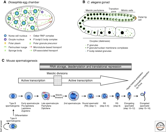
Germ granules during gametogenesis. (A) Transport of germ granule material in a Drosophila egg chamber. Various germ granule RNPs intermingle in the nurse cells’ cytoplasm. Green dashed lines represent migration of sponge bodies, red lines indicate the oskar RNP transport pathway. (Panel A adapted from Jaglarz et al. [2011] and reproduced, with permission, from Springer © 2011.) (B) Germ granules in the germline of an adult C. elegans hermaphrodite. Germ cells progress along an “assembly line” of development from mitotic stem cells at the distal end to the oocytes at the proximal end. Germ cells share a common cytoplasm, and are individualized late in oogenesis. P granules (green) are docked at the germ cell nuclei until diplotene, and transition to the cytoplasm along with nuclear pore complexes (green/black). P body-related granules (blue) accumulate in the shared cytoplasm (rachis) and in oocytes. (C) Mammalian spermatogenesis. Intermitochondrial cement (yellow dots) is present in type A spermatogonia, and disappears in type B spermatogonia until meiosis. The chromatoid body (CB; red filaments then becoming dots) appears in late pachytene cells as thick cytoplasmic fibers. After the first meiotic division, the CB starts condensing as one or two round foci. Except during meiotic divisions, transcription is active until the elongating spermatid stage, and many transcripts are translationally silenced and located in the CB. In elongated spermatids, transcripts are released from translational arrest.
References
-
- Anderson P, Kedersha N 2009. RNA granules: Post-transcriptional and epigenetic modulators of gene expression. Nat Rev Mol Cell Biol 10: 430–436 - PubMed
-
- Andonov MD, Chaldakov GN 1989. Morphological evidence for calcium storage in the chromatoid body of rat spermatids. Experientia 45: 377–378 - PubMed
Publication types
MeSH terms
Substances
Grants and funding
LinkOut - more resources
Full Text Sources
Molecular Biology Databases