. 2011 Aug 1;10(15):2521-8.
doi: 10.4161/cc.10.15.16584. Epub 2011 Aug 1.

Understanding the metabolic basis of drug resistance: therapeutic induction of the Warburg effect kills cancer cells

Affiliations

Understanding the metabolic basis of drug resistance: therapeutic induction of the Warburg effect kills cancer cells

Ubaldo E Martinez-Outschoorn et al. Cell Cycle. .

Abstract

Previously, we identified a form of epithelial-stromal metabolic coupling, in which cancer cells induce aerobic glycolysis in adjacent stromal fibroblasts, via oxidative stress, driving autophagy and mitophagy. In turn, these cancer-associated fibroblasts provide recycled nutrients to epithelial cancer cells, "fueling" oxidative mitochondrial metabolism and anabolic growth. An additional consequence is that these glycolytic fibroblasts protect cancer cells against apoptosis, by providing a steady nutrient stream of to mitochondria in cancer cells. Here, we investigated whether these interactions might be the basis of tamoxifen-resistance in ER(+) breast cancer cells. We show that MCF7 cells alone are Tamoxifen-sensitive, but become resistant when co-cultured with hTERT-immortalized human fibroblasts. Next, we searched for a drug combination (Tamoxifen + Dasatinib) that could over-come fibroblast-induced Tamoxifen-resistance. Importantly, we show that this drug combination acutely induces the Warburg effect (aerobic glycolysis) in MCF7 cancer cells, abruptly cutting off their ability to use their fuel supply, effectively killing these cancer cells. Thus, we believe that the Warburg effect in tumor cells is not the "root cause" of cancer, but rather it may provide the necessary clues to preventing chemo-resistance in cancer cells. Finally, we observed that this drug combination (Tamoxifen + Dasatinib) also had a generalized anti-oxidant effect, on both co-cultured fibroblasts and cancer cells alike, potentially reducing tumor-stroma co-evolution. Our results are consistent with the idea that chemo-resistance may be both a metabolic and stromal phenomenon that can be overcome by targeting mitochondrial function in epithelial cancer cells. Thus, simultaneously targeting both (1) the tumor stroma and (2) the epithelial cancer cells, with combination therapies, may be the most successful approach to anti-cancer therapy. This general strategy of combination therapy for overcoming drug resistance could be applicable to many different types of cancer.


Figures

Figure 1
Figure 1
Tamoxifen-induced apoptosis in MCF7 cells cultured alone: Dasatinib has no synergistic effect. MCF7 breast cancer cells were cultured alone, in the absence of fibroblasts and subjected to drug treatment with Tamoxifen (T; 12 µM) or Dasatinib (D; 2.5 nM), individually or in combination. Cell death in MCF7 cells was quantitated by FACS analysis via Annexin-V and PI staining. Note that Tamoxifen (T) treatment induces significant cell death in MCF7 cells. However, Dasatinib (D) has little or no appreciable effect. Tamoxifen plus Dasatinib (T + D) does not have a synergistic effect. In (A), “cell death” represents the percentage of cells that were Annexin-V(+) and/or PI(+), the sum of all three quadrants. In (B), only the Annexin-V(+) positivity is shown, yielding similar results. CTRL (control), represents MCF7 cells alone, treated with vehicle alone.
Figure 2
Figure 2
MCF7 cells co-cultured with fibroblasts are Tamoxifen-resistant: Dasatinib overcomes Tamoxifen-resistance. MCF7 breast cancer cells were co-cultured with fibroblasts, and then subjected to drug treatment with Tamoxifen (T; 12 µM) or Dasatinib (D; 2.5 nM), individually or in combination. Cell death in MCF7 cells was quantitated by FACS analysis via Annexin-V and PI staining. Note that individually Tamoxifen (T) or Dasatinib (D) treatment has little or no effect on MCF7 cell apoptosis. However, Tamoxifen plus Dasatinib (T + D) does have a clear synergistic effect, significantly killing MCF7 cells in co-culture. In (A), “cell death” represents the percentage of cells that were Annexin-V(+) and/or PI(+), the sum of all three quadrants. In (B), only the Annexin-V(+) positivity is shown, yielding similar results. CTRL (control), represents co-cultured MCF7 cells, treated with vehicle alone.
Figure 3
Figure 3
Dasatinib prevents a loss of Cav-1 expression in cancer-associated fibroblasts. MCF7 cells were co-cultured with fibroblasts, in the presence or absence of Dasatinib (2.5 or 5 nM). Then, these co-cultures were immuno-stained with antibody probes directed against Cav-1 (to detect fibroblasts) and Keratin-8/18 (to detect MCF7 epithelial cancer cells). Nuclei were visualized via DAPI staining. Note that Dasatinib treatment rescues the expression of Cav-1, that is normally downregulated in fibroblasts by co-culture with MCF7 cells. Similar results were obtained with both 2.5 and 5 nM Dasatinib. Control represents MCF7-co-cultures treated with vehicle alone.
Figure 4
Figure 4
Dasatinib has no effect on Cav-1 levels in stromal fibroblasts cultured alone. As in Figure 3, except that fibroblasts were cultured alone, in the absence of MCF7 cancer cells. Note that Dasatinib treatment does not affect expression of Cav-1. Control represents fibroblasts alone treated with vehicle alone.
Figure 5
Figure 5
Dasatinib preferentially affects fibroblasts in co-culture. MCF7 breast cancer cells were co-cultured with fibroblasts, and then subjected to drug treatment with Tamoxifen (T; 12 µM) or Dasatinib (D; 2.5 nM), individually or in combination. Cell death in fibroblasts was quantitated by FACS analysis via Annexin-V and PI staining. Note that Dasatinib (D) preferentially affects fibroblasts in co-culture, while Tamoxifen (T) has only minor effects. In (A), “cell death” represents the percentage of cells that were Annexin-V(+) and/or PI(+), the sum of all three quadrants. In (B), only the Annexin-V(+) positivity is shown, yielding similar results. CTRL (control), represents co-cultured fibroblasts, treated with vehicle alone.
Figure 6
Figure 6
Tamoxifen plus Dasatinib increases glucose uptake in MCF7 cells co-cultured with fibroblasts. MCF7 breast cancer cells were co-cultured with fibroblasts, and then subjected to drug treatment with Tamoxifen (T; 12 µM) or Dasatinib (D; 2.5 nM), individually or in combination. Glucose uptake in MCF7 cells was quantitated by FACS analysis, using NBD-2-deoxy-glucose as a probe. Tamoxifen plus Dasatinib (T + D) has a clear synergistic effect, significantly increasing glucose uptake nearly 4-fold in MCF7 cells in co-culture. CTRL (control), represents co-cultured MCF7 cells, treated with vehicle alone.
Figure 7
Figure 7
Tamoxifen plus Dasatinib normalizes the glucose uptake ratio in co-cultures (Fibroblasts/MCF7 Cells). As in Figure 6, except that glucose uptake was measured in both co-cultured MCF7 cells and fibroblasts. Note that Tamoxifen plus Dasatinib (T + D) has a clear synergistic effect, significantly normalizing the glucose uptake ratio (Fibro/MCF7) from nearly 2, to less than 1.
Figure 8
Figure 8
Tamoxifen plus Dasatinib reduces mitochondrial activity in MCF7 cells co-cultured with fibroblasts. MCF7 breast cancer cells were co-cultured with fibroblasts, and then subjected to drug treatment with Tamoxifen (T; 12 µM) or Dasatinib (D; 2.5 nM), individually or in combination. Mitochondrial activity in MCF7 cells was quantitated by FACS analysis, using MitoTracker as a probe. Tamoxifen plus Dasatinib (T + D) has a clear synergistic effect, significantly decreasing mitochondrial activity >six-fold in MCF7 cells in co-culture. CTRL (control), represents co-cultured MCF7 cells, treated with vehicle alone.
Figure 9
Figure 9
Tamoxifen plus Dasatinib normalizes the mitochondrial activity ratio in co-cultures (MCF7 cells/fibroblasts). As in Figure 8, except that mitochondrial activity was measured in both co-cultured MCF7 cells and fibroblasts. Note that Tamoxifen plus Dasatinib (T + D) has a synergistic effect, significantly normalizing the mitochondrial activity ratio (MCF7/Fibro) from greater than 2.5, to less than 1.5.
Figure 10
Figure 10
Tamoxifen plus Dasatinib reduces mitochondrial activity in fibroblasts co-cultured with MCF7 cells. MCF7 breast cancer cells were co-cultured with fibroblasts, and then subjected to drug treatment with tamoxifen (T; 12 µM) or Dasatinib (D; 2.5 nM), individually or in combination. Mitochondrial activity in fibroblasts was then quantitated by FACS analysis, using Mitotracker as a probe. Tamoxifen plus Dasatinib (T + D) has a clear synergistic effect, significantly decreasing mitochondrial activity nearly four-fold in fibroblasts in co-culture. CTRL (control), represents co-cultured fibroblasts, treated with vehicle alone.
Figure 11
Figure 11
Tamoxifen plus Dasatinib reduces ROS activity in MCF7 cells co-cultured with fibroblasts. MCF7 breast cancer cells were co-cultured with fibroblasts, and then subjected to drug treatment with Tamoxifen (T; 12 µM) or Dasatinib (D; 2.5 nM), individually or in combination. ROS production in MCF7 cells was then quantitated by FACS analysis, using DCF-DA as a probe. Tamoxifen plus Dasatinib (T + D) has a clear synergistic anti-oxidant effect, significantly decreasing ROS activity ∼three-fold in MCF7 cells in co-culture. CTRL (control), represents co-cultured MCF7 cells, treated with vehicle alone.
Figure 12
Figure 12
Tamoxifen plus Dasatinib reduces ROS activity in fibroblasts co-cultured with MCF7 cells. MCF7 breast cancer cells were co-cultured with fibroblasts, and then subjected to drug treatment with Tamoxifen (T; 12 µM) or Dasatinib (D; 2.5 nM), individually or in combination. ROS production in fibroblasts was then quantitated by FACS analysis, using DCF-DA as a probe. tamoxifen plus Dasatinib (T + D) has a clear synergistic anti-oxidant effect, significantly decreasing ROS activity ∼14-fold in fibroblasts in co-culture. CTRL (control), represents co-cultured fibroblasts, treated with vehicle alone.
Figure 13
Figure 13
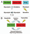
Understanding the metabolic basis of drug resistance: therapeutic induction of the Warburg Effect kills cancer cells. (A) In summary, our results indicate that MCF7 cells are sensitive to Tamoxifen (T), when they are cultured alone, and are metabolically glycolytic. However, MCF7 cells become resistant to Tamoxifen, when they are co-cultured with fibroblasts, and become oxidative (Upper). Thus, Tamoxifen-resistance may conferred by this metabolic shift in MCF7 cells from a glycolytic to an oxidative state, which is dependent on mitochondrial oxidative phosphorylation in cancer cells. As a consequence, we should be able to overcome Tamoxifen-resistance, by shifting MCF7 cells back to their glycolytic state. For this purpose, we developed a Tamoxifen-based drug combination that induces aerobic glycolysis (i.e., the Warburg Effect) in co-cultured MCF7 cells, resulting in their apoptotic cell death (Lower). As such, this drug combination (Tamoxifen + Dasatinib; T + D) is synthetically lethal with the “reverse Warburg effect” in ER(+) breast cancer cells. This new strategy for overcoming drug resistance should be generally applicable to many different types of cancer. The blue arrow indicates the direction of net energy flow, from fibroblasts to cancer cells, due to the “reverse Warburg effect.” (B) Targeting both the tumor stroma and epithelial cancer cells with a combination therapy. Cancer cells use oxidative stress as a “weapon” to extract recycled nutrients (via autophagy/mitophagy) and high-energy metabolites (via aerobic glycolysis) from adjacent stromal fibroblasts. Combination therapy with Tamoxifen plus Dasatinib may interrupt this vicious metabolic cycle, by simultaneously targeting both epithelial cancer cells and their “food supply,” the fibroblasts.

References

    1. Witkiewicz AK, Casimiro MC, Dasgupta A, Mercier I, Wang C, Bonuccelli G, et al. Towards a new “stromal-based” classification system for human breast cancer prognosis and therapy. Cell Cycle. 2009;8:1654–1658. doi: 10.4161/cc.8.11.8544. - DOI - PubMed
    1. Witkiewicz AK, Dasgupta A, Nguyen KH, Liu C, Kovatich AJ, Schwartz GF, et al. Stromal caveolin-1 levels predict early DCIS progression to invasive breast cancer. Cancer Biol Ther. 2009;8:1071–1079. doi: 10.4161/cbt.8.11.8874. - DOI - PubMed
    1. Witkiewicz AK, Dasgupta A, Sammons S, Er O, Potoczek MB, Guiles F, et al. Loss of stromal caveolin-1 expression predicts poor clinical outcome in triple negative and basal-like breast cancers. Cancer Biol Ther. 2010;10:135–143. doi: 10.4161/cbt.10.2.11983. - DOI - PMC - PubMed
    1. Witkiewicz AK, Dasgupta A, Sotgia F, Mercier I, Pestell RG, Sabel M, et al. An absence of stromal caveolin-1 expression predicts early tumor recurrence and poor clinical outcome in human breast cancers. Am J Pathol. 2009;174:2023–2034. doi: 10.2353/ajpath.2009.080873. - DOI - PMC - PubMed
    1. Witkiewicz AK, Kline J, Queenan M, Brody JR, Tsirigos A, Bilal E, et al. Molecular profiling of a lethal tumor microenvironment, as defined by stromal caveolin-1 status in breast cancers. Cell Cycle. 2011;10:1794–1809. - PMC - PubMed

Publication types

MeSH terms