. 2012 Jan;26(1):106-16.
doi: 10.1038/leu.2011.185. Epub 2011 Jul 19.

Conditioning for hematopoietic transplantation activates the complement cascade and induces a proteolytic environment in bone marrow: a novel role for bioactive lipids and soluble C5b-C9 as homing factors

Affiliations

Conditioning for hematopoietic transplantation activates the complement cascade and induces a proteolytic environment in bone marrow: a novel role for bioactive lipids and soluble C5b-C9 as homing factors

C H Kim et al. Leukemia. 2012 Jan.

Abstract

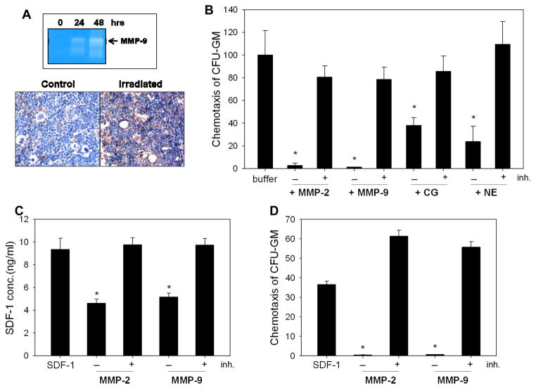
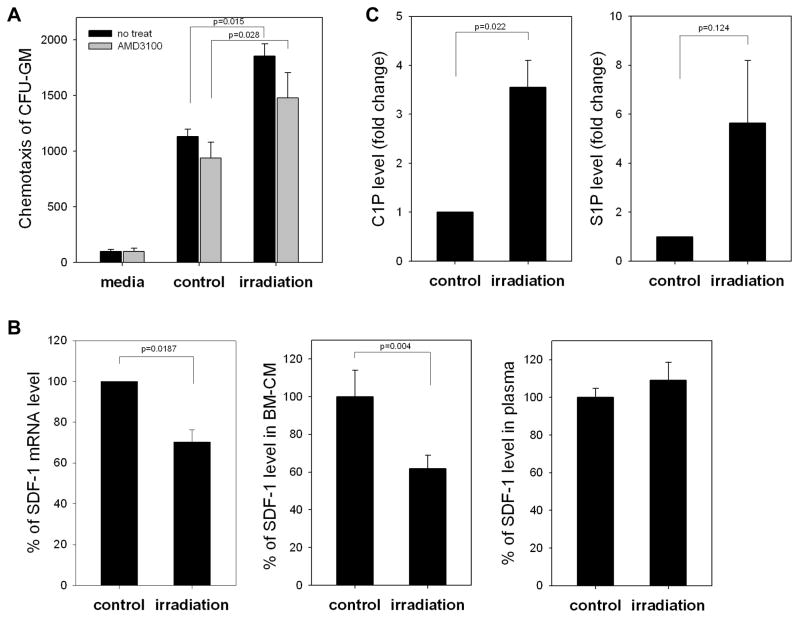
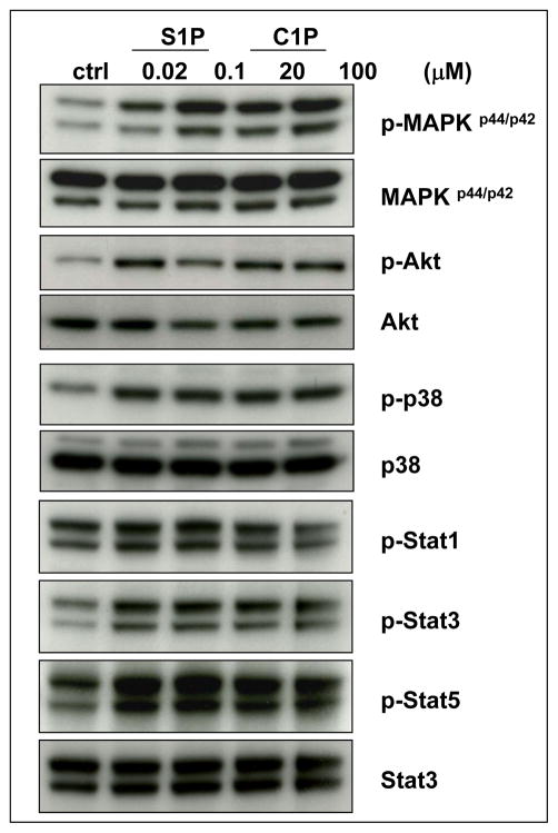
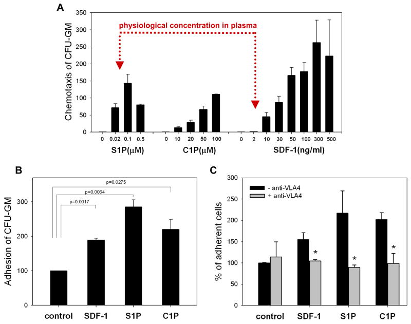
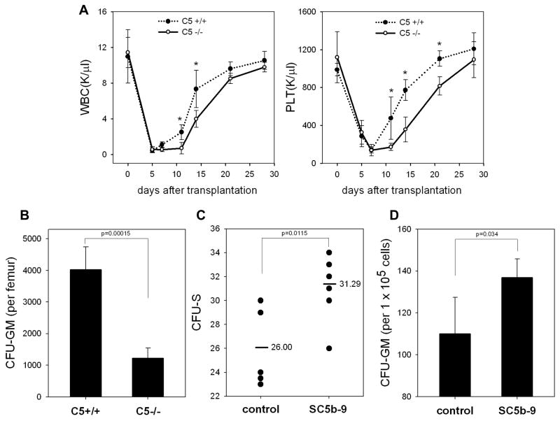
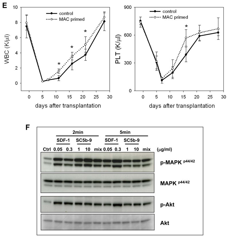
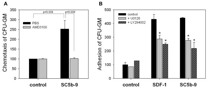
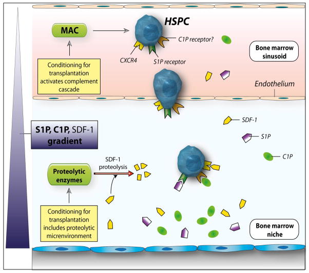
We have observed that conditioning for hematopoietic transplantation by lethal irradiation induces a proteolytic microenvironment in the bone marrow (BM) that activates the complement cascade (CC). As a result, BM is enriched for proteolytic enzymes and the soluble form of the terminal product of CC activation, the membrane attack complex C5b-C9 (MAC). At the same time, proteolytic enzymes induced in irradiated BM impair the chemotactic activity of α-chemokine stromal-derived factor-1 (SDF-1). As SDF-1 is considered a crucial BM chemoattractant for transplanted hematopoietic stem/progenitor cells (HSPCs), we sought to determine whether other factors that are resistant to proteolytic enzymes have a role in this process, focusing on proteolysis-resistant bioactive lipids. We found that the concentrations of sphingosine-1-phosphate (S1P) and ceramide-1-phosphate (C1P) increase in the BM after conditioning for transplantation and that both S1P and, as we show here for the first time, C1P are potent chemoattractants for HSPCs. Next, we observed that C5-deficient mice that do not generate MAC show impaired engraftment of HSPCs. In support of a role for MAC in homing and engraftment, we found that soluble MAC enhances in a CR3 (CD11b/CD18)-dependent manner the adhesion of HSPCs to BM stromal cells and increases the secretion of SDF-1 by BM stroma. We conclude that an increase in BM levels of proteolytic enzyme-resistant S1P and C1P and activation of CC, which leads to the generation of MAC, has an important and previously underappreciated role in the homing of transplanted HSPCs.


Figures

Figure 1
Figure 1. Myeloablative conditioning for hematopoietic transplantation by lethal irradiation induces a proteolytic microenvironment in murine BM
Panel A: Zymography revealed that the activity of MMP-9 is increased at 24 and 48 hours in conditioned media harvested from BM cells after lethal (1000 cGy) γ-irradiation (upper panel). The complement cascade becomes activated and MAC is deposited in the BM microenvironment of mice that were conditioned for transplantation by lethal γ-irradiation (lower panel). Because it is a peptide, SDF-1 was disrupted by proteolytic enzymes that are elevated in irradiated BM. Panel B: Chemotactic activity of SDF-1 against clonogenic progenitors decreases after exposure to MMP-2, MMP-9, cathepsin G (CG), or neutrophil elastase (NE). Disruption of SDF-1 was prevented by addition of protease-specific inhibitors. Panel C: SDF-1 was exposed to MMP-2 (100ng) or MMP-9 (100ng) with or without inhibitor (ARP101, 100ng) pre-incubation, and SDF-1 concentration was evaluated by ELISA. Panel D: The SDF-1 samples from Panel C were tested for chemotactic activity against BM-derived CFU-GM. The data shown in panels B–D represent the combined results from three independent experiments carried out in triplicate per group. * p<0.0001.
Figure 2
Figure 2. SDF-1 level decreases in irradiated bone marrow (BM)
Panel A: Conditioned media from irradiated BM cells enhance chemotactic activity of normal clonogeneic progenitors (black bars), but this effect was partially disrupted by blocking CXCR4 with AMD3100 (1μM, gray bars). Panel B: SDF-1 at both the mRNA level (real-time PCR, left panel) and protein level (ELISA, middle panel) decreases after 24 hours in lethally irradiated BM (1000 cGy). At the same time, we did not observe changes in the SDF-1 level in PB of irradiated mice (right panel). Panel C: Mass spectrometry analysis shows that ceramide-1-phosphate (C1P) and sphingosine-1-phosphate (S1P) become upregulated in murine BM after conditioning for hematopoietic transplantation by lethal irradiation. The data shown in panels A–C represent the combined results from three independent experiments carried out in triplicate per group.
Figure 3
Figure 3. Bioactive lipids stimulate signaling pathways in murine Sca-1+ HSPCs
C1P and S1P activate several signaling pathways in murine BMMNC that are crucial for cell migration and adhesion: MAPKp44/42, Akt, p38, Stat-3, and Stat-5. Before stimulation, cells were starved overnight in RPMI containing 0.5% BSA in an incubator and subsequently stimulated with S1P (0.02μM or 0.1μM) or C1P (20μM or 100μM) for 5 min. Experiments were repeated independently three times with similar results. A representative western blot is shown.
Figure 4
Figure 4. Bioactive lipids show strong chemotactic activity against HSPCs
Panel A: The chemotatic dose response of S1P, C1P, and SDF-1 against murine clonogenic progenitors is shown. Note that the physiological concentration of SDF-1 in biological fluids (e.g., serum) is approximately 2–5ng/ml, and at this concentration SDF-1 is not effective as a chemoattractant. In contrast, S1P is already a strong chemoattractant for murine BM-derived hematopoietic progenitors at biologically relevant concentrations. Panel B: Both C1P and S1P, like SDF-1, increase adhesion of murine clonogenic progenitors (Sca-1+ cells) to BM-derived stromal cells. The data shown represent the combined results from three independent experiments carried out in triplicate per group. Panel C: In blocking studies, Sca-1+ cells were incubated with 10μg/ml anti-VLA4 integrin mAb for 30 minutes prior to VCAM-1 adhesion assay. The data shown represent the combined results from two independent experiments carried out in quadruplicate per group. * p<0.05.
Figure 5
Figure 5. C5-deficient mice show an engraftment defect in transplanted HSPCs
Panel A: Wild type (C5+/+) and C5-deficient (C5−/−) animals, which do not generate MAC, were lethally irradiated and transplanted with wild type bone marrow mononuclear cells. We followed the subsequent recovery of hematopoietic parameters in these animals. C5−/− mice have recovery of peripheral blood leukocytes (upper panel) and platelets (lower panel) delayed by 3–5 days. The data shown represent the combined results from two independent experiments performed with 10 mice/group. * p<0.001. Panel B: By day 12, there is a decrease in the number of CFU-GM in BM of C5−/− mice transplanted with wt BM cells. Panel C: The number of CFU-S colonies formed in the spleens of wt mice were counted after transplantation of wt BMMNC cells, which were pre-stimulated or unstimulated before transplantation by sC5b-9 (10μg/ml), and injected intravenously into lethally irradiated wild type animals. Twelve days later, spleen was isolated and the number of CFU-S evaluated. Panel D: The number of CFU-GM progenitors in BM of wt transplanted mice with SC5b-9-primed or unprimed BM cells. The data shown in panels C and D represent the combined results from three independent experiments carried out in triplicate per group (n=9). Panel E: Wild type mice were lethally irradiated and transplanted with wild type BMMNC after SC5b-9 treatment (primed) or not (unprimed control). We followed the subsequent recovery of hematopoietic parameters in these animals. Mice that were transplanted with SC5b-9-primed BMMNCs have a much faster recovery rate for peripheral blood leukocytes (left panel) and platelets (right panel). * p<0.05. Panel F: Soluble MAC, like SDF-1, activates MAPKp44/42 and Akt in normal murine Sca-1+ cells. “Mix” indicates SDF-1 (0.05μg/ml) plus SC5b-9 (1μg/ml). Experiments were repeated independently three times with similar results. A representative western blot is shown.
Figure 6
Figure 6. MAC affects several steps crucial for stem cell homing
Panel A: Normal murine BM-derived stromal cells were incubated with soluble MAC (sC5b-9, 10μg/ml for 3 hrs) and subsequently conditioned media were harvested from these cells and assayed for their chemotactic activity against normal murine hematopoietic progenitors (black bars). Importantly, this chemotactic effect was totally inhibited after blockage of the CXCR4 receptor by AMD3100 treatment (gray bars). Panel B: Soluble MAC (sC5b-9), like SDF-1, strongly increases adhesiveness of murine hematopoietic progenitor cells (Sca-1+) to murine bone marrow-derived stroma. In inhibition studies, Sca-1+ cells were treated with MAPK inhibitor (U0126, 10μM) or Akt inhibitor (LY294002, 20μM) for 1 h before adhesion assay. The data shown represent the combined results from three independent experiments carried out in triplicate per group.
Figure 7
Figure 7. The involvement of bioactive lipids in homing and engraftment of HSPCs
Conditioning for transplantation by radio-chemotherapy induces a proteolytic microenvironment in BM and activates the complement cascade, which leads to generation of soluble MAC. Several proteolytic enzymes are released that decrease the SDF-1 level in BM. At the same time, BM cells damaged by conditioning for transplantation by lethal irradiation release C1P and S1P, which are bioactive lipids resistant to proteolytic enzymes. As potent chemoattractants, both play an important role in homing of stem cells to BM. In addition, MAC generated by CC activation enhances adhesiveness of HSPCs to BM stroma and secretion of SDF-1 by these cells. This increase in SDF-1 secretion may somehow ameliorate the drop in SDF-1 level in such a highly proteolytic microenvironment. What is not shown in this scheme is that CC cleavage fragments, such as C3a and iC3b, also contribute to the homing of HSPCs, as reported by us in the past , . By increasing the PGE2 level in BM, these CC cleavage fragments level may also modulate the seeding of HSPCs in BM, in addition to the bioactive lipids C1P and S1P.

References

    1. Levesque JP, Takamatsu Y, Nilsson SK, Haylock DN, Simmons PJ. Vascular cell adhesion molecule-1 (CD106) is cleaved by neutrophil proteases in the bone marrow following hematopoietic progenitor cell mobilization by granulocyte colony-stimulating factor. Blood. 2001;98:1289–1297. - PubMed
    1. Peled A, Grabovsky V, Habler L, Sandbank J, Arenzana-Seisdedos F, Petit I, et al. The chemokine SDF-1 stimulates integrin-mediated arrest of CD34+ cells on vascular endothelium under shear flow. J Clin Invest. 1999;104:1199–1211. - PMC - PubMed
    1. Ratajczak MZ, Reca R, Wysoczynski M, Kucia M, Baran JT, Allendorf DJ, et al. Transplantation studies in C3-deficient animals reveal a novel role of the third complement component (C3) in engraftment of bone marrow cells. Leukemia. 2004;18:1482–1490. - PubMed
    1. Cho SY, Xu M, Roboz J, Lu M, Mascarenhas J, Hoffman R. The effect of CXCL12 processing on CD34+ cell migration in myeloproliferative neoplasms. Cancer Res. 2010;70:3402–3410. - PubMed
    1. Lévesque JP, Hendy J, Takamatsu Y, Simmons PJ, Bendall LJ. Disruption of the CXCR4/CXCL12 chemotactic interaction during hematopoietic stem cell mobilization induced by GCSF or cyclophosphamide. J Clin Invest. 2003;111:187–196. - PMC - PubMed

Publication types