. 2011 Jun 15;49(12):832-864.
doi: 10.1002/polb.22259.

Biomedical Applications of Biodegradable Polymers

Affiliations

Biomedical Applications of Biodegradable Polymers

Bret D Ulery et al. J Polym Sci B Polym Phys. .

Abstract

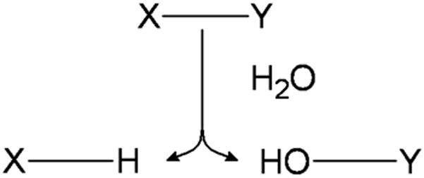
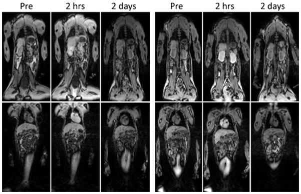
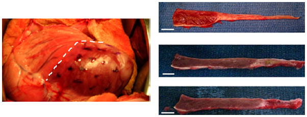
Utilization of polymers as biomaterials has greatly impacted the advancement of modern medicine. Specifically, polymeric biomaterials that are biodegradable provide the significant advantage of being able to be broken down and removed after they have served their function. Applications are wide ranging with degradable polymers being used clinically as surgical sutures and implants. In order to fit functional demand, materials with desired physical, chemical, biological, biomechanical and degradation properties must be selected. Fortunately, a wide range of natural and synthetic degradable polymers has been investigated for biomedical applications with novel materials constantly being developed to meet new challenges. This review summarizes the most recent advances in the field over the past 4 years, specifically highlighting new and interesting discoveries in tissue engineering and drug delivery applications.


Figures

Figure 1
Figure 1
The hydrolytically sensitive bond X-Y is cleaved by a water molecule yielding the products of X-H and HO-Y.
Figure 2
Figure 2
Conversion of CT images into micro- and nanostructure controlled PLA scaffolds. A CT image of a hand (left) with a non-traditional defect (shown in purple) is converted into a wax mold which can be filled with PLA to create a scaffold with controllable pore size on the micro scale (center) and fiber size on the nano scale (right). (reprinted from with permission from Elsevier.)
Figure 3
Figure 3
PLGA scaffolds with villi architecture generated by indirect three-dimensional printing with villus diameter, height and intervillus spacing of (a) 0.5, 1, 0.5 mm; (b) 0.5, 1, 1 mm; (c) 1, 1, 1 mm, respectively. (reprinted from with permission from Wiley.)
Figure 4
Figure 4
Cylindrical porous poly(L-lactide-co-caprolactone) scaffold loaded with fibrochondryocytes (fibrocartilage sections) on either end with fibroblasts (ligament section) in the center in order to mimic the ligament-bone interfacial tissues. (reprinted from with permission from Elsevier.)
Figure 5
Figure 5
In vitro release of bovine serum albumin from poly(sebacic anhydride-co-1,6-bis-p-carboxyphenoxy hexane) microparticles in phosphate-buffered saline (pH 7.4). (reprinted from with permission from Elsevier.)
Figure 6
Figure 6
In vitro hydrolysis of poly(cyclohexane-1,4-diyl acetone dimethyl ketal) is greatly influenced by surrounding pH evidenced by its half-life of 24.1 days in pH 4.5 and 4 years in pH 7.4. (reprinted from with permission from the American Chemical Society.)
Figure 7
Figure 7
Optical microscopy image of a polyphosphazene coated metallic microneedle (left) and histological section of porcine skin after coated microneedle insertion (right). (reprinted from )
Figure 8
Figure 8
Photomicrographs of keratinocyte-fibroblast co-culture engineered skin tissue with the addition of collagen microsphere supported sweat gland cell constructs. (A) Hemotoxylin and Eosin (H&E) staining after two weeks of in vitro co-cultivation showed differentiated tissue layers (E: epithelium and D: dermis) with a bud-like structure (black arrow) where sweat gland constructs were loaded. (B) H&E staining of six week post-implantation in vivo skin tissue showed the continued presence of bud-like structures in the dermis layer (black arrow). (C) Fluorescence microscope observation showed DiO-positive cells (green) confirming the presence of still viable sweat glands. (reprinted from with permission from Elsevier.)
Figure 9
Figure 9
Magnetic resonance imaging of rhesus monkeys before contrast injection and at 2 h and 2 days after injection of Magnevist at 0.1 mmol Gd/kg (left) and PG-Gd 0.01 mmol Gd/kg (right). Enhancements of blood vessel, heart, kidney and liver are clearly visualized at 2 h after PG-Gd injection at a tenth of the dose of Magnevist. By 2 days, the contrast agent has been mostly cleared with both contrast agents. (reprinted from with permission from Wiley.)
Figure 10
Figure 10
MeHA injection into a sheet heart after infarction helps prevent subsequent myocardial damage. Multiple injections (dots) of MeHA were given into the infarcted area (right of dashed line) (left). Representative images of myocardial wall thickness 8 weeks post treatment with no scaffold (top right), MeHA High scaffold (middle right), and MeHA Low scaffold (bottom right). (reprinted from )

References

    1. Williams DF. Biomaterials. 2009;30:5897–5909. - PubMed
    1. Williams DF. The Williams Dictionary of Biomaterials. Liverpool University Press; Liverpool: 1999.
    1. Schmitt EE, Polistina RA USPTO. United States; 1967.
    1. Schmitt EE, Polistina RA USPTO. United States; 1963.
    1. Ibim SEM, Ambrosio AMA, Kwon MS, El-Amin SF, Allcock HR, Laurencin CT. Biomaterials. 1997;18:1565–1569. - PubMed

LinkOut - more resources