. 2011:5:80-95.
doi: 10.2174/1874357901105010080. Epub 2011 Jul 4.

Small molecule inhibitors of human papillomavirus protein - protein interactions

Affiliations

Small molecule inhibitors of human papillomavirus protein - protein interactions

C M D'Abramo et al. Open Virol J. 2011.

Abstract

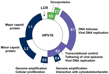
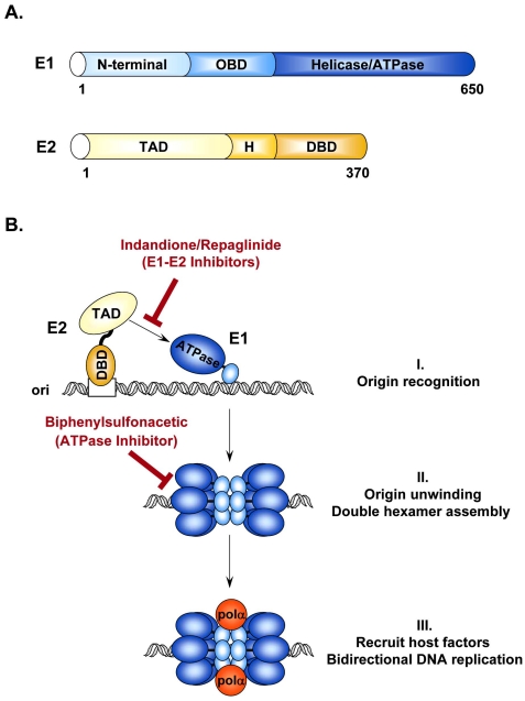
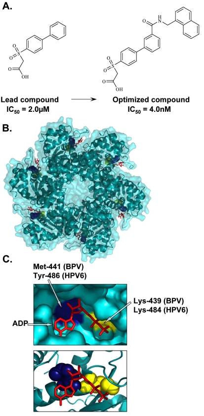
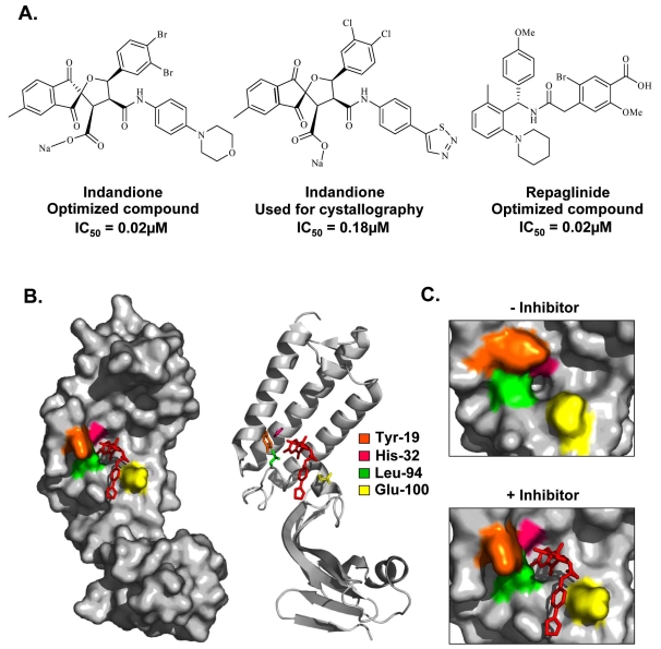
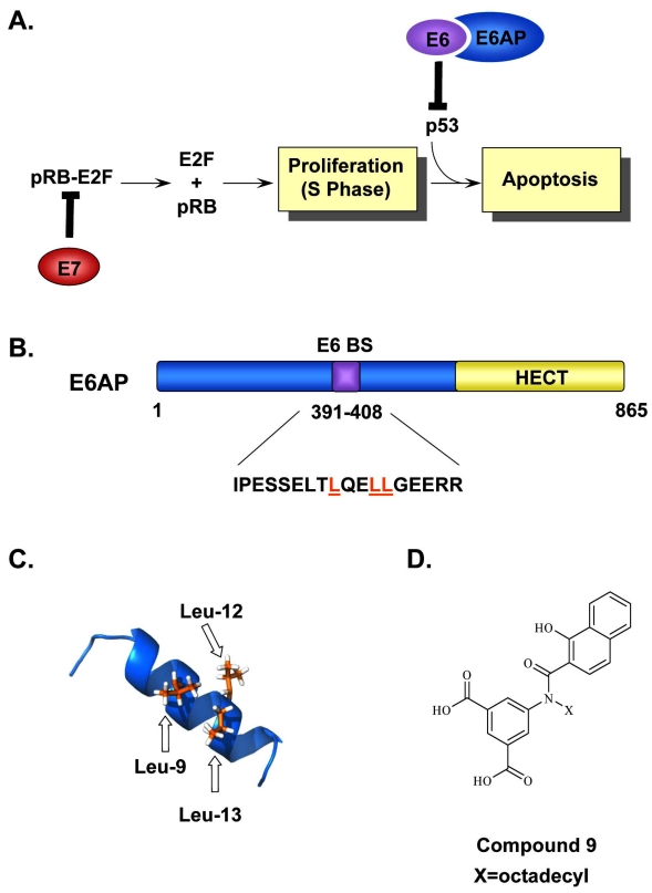
Human papillomaviruses (HPV) have now been identified as a necessary cause of benign and malignant lesions of the differentiating epithelium, particularly cervical cancer, the second most prevalent cancer in women worldwide. While two prophylactic HPV vaccines and screening programs are available, there is currently no antiviral drug for the treatment of HPV infections and associated diseases. The recent progress toward the identification and characterization of specific molecular targets for small molecule-based approaches provides prospect for the development of effective HPV antiviral compounds. Traditionally, antiviral therapies target viral enzymes. HPV encode for few proteins, however, and rely extensively on the infected cell for completion of their life cycle. This article will review the functions of the viral E1 helicase, which encodes the only enzymatic function of the virus, of the E2 regulatory protein, and of the viral E6 and E7 oncogenes in viral replication and pathogenesis. Particular emphasis will be placed on the recent progress made towards the development of novel small molecule inhibitors that specifically target and inhibit the functions of these viral proteins, as well as their interactions with other viral and/or cellular proteins.

Keywords: E1; E2; E6; E6AP.; HPV; cervical cancer; protein interaction; small molecule inhibitor.


Figures

Fig. (1)
Fig. (1)
Genomic organization of the HPV genome. Schematic representation of the HPV16 circular genome showing the location of the early (E) and late genes (L1 and L2), and of the long control region (LCR). The HPV genome encodes eight well-characterized proteins, whose functions are indicated. Among them are the viral replication proteins E1 and E2 (violet) and the viral oncogenes E6 and E7 (green), all of which have been validated as essential for viral pathogenesis and represent genuine targets for small molecule-based approaches for the treatment of HPV-associated diseases.
Fig. (2)
Fig. (2)
Initiation of HPV DNA replication. (A) Schematic representation of the viral proteins E1 and E2 required for replication of the HPV genome. E1 and E2 are approximately 650 and 370 amino acids in length, respectively. Locations of the different functional domains in each protein are indicated. OBD: origin binding domain; TAD: transactivation domain; H: hinge region; DBD: DNA-binding domain. (B) Schematic diagram of the initiation of HPV DNA replication. (I) Replication is initiated by the recruitment of E1 (blue), by E2 (yellow), to the viral origin. This recruitment step involves an essential protein-protein interaction between the TAD of E2 and the helicase domain of E1 that can be antagonized by the Indandione or Repaglinide class of small molecule inhibitors. (II) E2 recruits additional E1 molecules and promotes their assembly into a replication-competent double hexameric helicase. ATP also stimulates the oligomerization of E1 and is further needed to power the helicase activity of E1. Biphenylsulfonacetic acid inhibitors have been identified that abrogate the ATPase and helicase activities of E1. (III) Finally, E1 interacts with host cell replication factors such as polymerase α primase (pol α; orange) to promote bidirectional replication of the viral genome.
Fig. (3)
Fig. (3)
Inhibition of E1 ATPase activity. (A) Structures of the lead and optimized biphenylsulfonacetic acid inhibitors of the E1 ATPase activity with IC50 values of 2.0µM and 4.0nM, respectively. (B) Crystal structure of the hexameric C-terminal helicase domain of bovine papillomavirus (BPV) E1 (PDB accession number 2GXA [173]). (C) Enlarged views of the ATP-binding pocket displaying the locations of the highly conserved catalytic Lys-439 (yellow), essential for ATP interaction and catalysis, and Met-441, (blue) important for the activity of biphenylsulfonacetic acid inhibitors. These residues are equivalent to Lys-484 and Tyr-486, respectively, in HPV6 E1. Bound ADP (red) is depicted in stick representation.
Fig. (4)
Fig. (4)
Inhibition of the E1-E2 protein interaction. (A) Structures and potencies of optimized indandione and repaglinide inhibitors of the E1-E2 protein-protein interaction. (B) Surface and ribbon representation of the HPV11 E2 TAD-indandione inhibitor complex (PDB accession number 1R6K [101]). The structure of the inhibitor used for crystallography is shown in (A). The amino acid residues that form the hydrophobic inhibitor-binding pocket are depicted in stick representation and are colored according to the legend in the figure. (C) Enlarged view of the hydrophobic pocket in the absence (left panel) and presence (right panel) of inhibitor, displaying the significant movement of the amino acids Tyr-19, His-32, Leu-94, and Glu-100 upon inhibitor binding.
Fig. (5)
Fig. (5)
Inhibition of the E6-E6AP interaction. (A) Simplified model of how the HPV oncogenes E6 and E7 stimulate cellular proliferation. Binding of E7 to pRb leads to the release and activation of the E2F transcription factors and drives differentiating keratinocytes into S-phase. This unscheduled DNA synthesis triggers a p53-dependent cell cycle arrest and apoptotic response that is prevented by E6, through its interaction with E6AP, and targets p53 for proteasomal degradation. (B) Schematic representation of E6AP. E6AP possesses several well-characterized functional domains including a HECT domain (yellow) and an E6-binding site (E6BS). The amino acid sequence corresponding to the E6BS is indicated and the three conserved leucine residues, Leu-9, Leu-12, Leu-13, are highlighted in orange. (C) NMR structure of the E6AP peptide showing the positions of the three leucine residues, Leu-9, Leu-12, and Leu-13 important for E6 binding are colored in orange (PDB accession number 1EQX [152]). (D) Structure of Compound 9, an E6-E6AP inhibitor used as a starting point for the synthesis of other closely related inhibitors of E6 activity.

References

    1. de Villiers EM, Fauquet C, Broker TR, Bernard HU, zur Hausen H. Classification of papillomaviruses. Virology. 2004;324:17–27. - PubMed
    1. Munger K, Baldwin A, Edwards KM, et al. Mechanisms of human papillomavirus-induced oncogenesis. J Virol. 2004;78:11451–60. - PMC - PubMed
    1. Woodman CB, Collins SI, Young LS. The natural history of cervical HPV infection: unresolved issues. Nat Rev Cancer. 2007;7:11–22. - PubMed
    1. Gillison ML. Human papillomavirus-associated head and neck cancer is a distinct epidemiologic, clinical, and molecular entity. Semin Oncol. 2004;31:744–54. - PubMed
    1. Syrjanen S. Human papillomavirus (HPV) in head and neck cancer. J Clin Virol. 2005;32(Suppl 1):S59–66. - PubMed

Grants and funding

LinkOut - more resources