

Case Reports
doi: 10.4081/rt.2011.e20.
Intracerebral metaplastic meningioma with prominent ossification and extensive calcification
Affiliations
- PMID: 21769319
- PMCID: PMC3132124
- DOI: 10.4081/rt.2011.e20
Item in Clipboard
Case Reports
Intracerebral metaplastic meningioma with prominent ossification and extensive calcification
Rare Tumors.
.
Abstract
We present a patient (male 26 years) with a short history of recurrent seizures induced by a largely intracerebrally located frontal lobe meningioma. The tumor displayed a heretofore unpublished combination of extensive metaplastic bone formation and prominent non-psammomatous calcifications with focal chicken-wire pattern.
Keywords: brain; calcification.; meningioma; metaplastic; ossification.
Figures
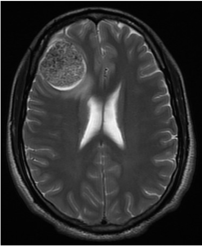
A right frontal lobe tumor. Magnetic resonance imaging scan of the brain was performed and revealed a 4.0− cm calcified tumor associated with edema of the adjacent brain parenchyma.
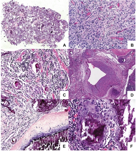
A heterogeneous tumor with areas of calcification and bone formation. (A) Tumor seen at low power showing cellular areas with areas of calcification and ossification. (B) The cellular areas are composed of plump, elongated cells with meningothelial features. (C) Prominent lymphoplasmacytic infiltrate is present within the cellular areas. (D) The highly vascularized tumour also contain dysplastic blood vessels. (E) The areas of calcification show crystalline deposits of calcium merge seamlessly with woven bone. No osteoblastic rimming is seen. (F) An interlacing chicken-wire-like calcification is seen in several areas.
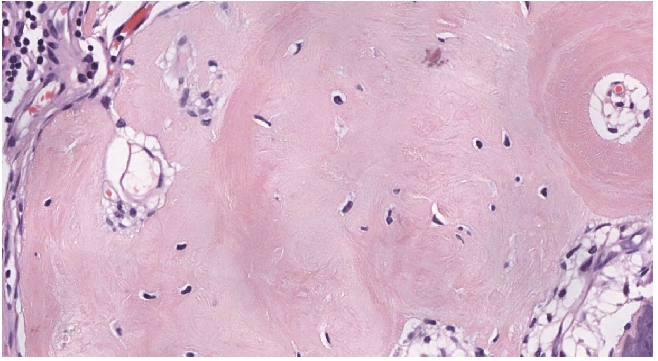
Bone formation within the tumour. Bony trabeculae with no osteoblastic rimming is seen within the tumour.
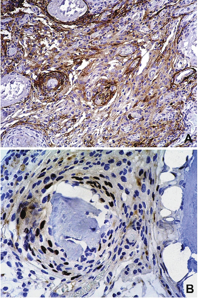
The tumor expresses epithelial membrane antigen and progesterone receptor. Sections of the tumor were stained with anti-EMA and anti-progesterone receptor primary antibodies and couterstained with hematoxylin. (A) Diffuse expression of cytoplasmic EMA is seen. (B) Scattered cells show nuclear expression of progesterone receptor.
References
-
- Roncaroli F, Scheithauer BW, Laeng RH, et al. Lipomatous meningioma: a clinicopathologic study of 18 cases with special reference to the issue of metaplasia. Am J Surg Pathol. 2001;25:769–75. - PubMed
-
- Uchida K, Nakajima H, Yayama T, et al. Immunohistochemical findings of multiple ossified en plaque meningiomas in the thoracic spine. J Clin Neurosci. 2009;16:1660–2. - PubMed
-
- Kato K, Chernov M, Urino T, et al. Ossified frontosphenoorbital meningioma en plaque, mimicking extensive hyperostosis. Minim Invasive Neurosurg. 2008;51:237–9. - PubMed
-
- Liu CL, Lai PL, Jung SM, Liao CC. Thoracic ossified meningioma and osteoporotic burst fracture: treatment with combined vertebroplasty and laminectomy without instrumentation: case report. J Neurosurg Spine. 2006;4:256–9. - PubMed
Publication types
LinkOut - more resources
Full Text Sources
