. 2011 Nov;26(5):1127-32.
doi: 10.3892/or.2011.1395. Epub 2011 Jul 18.

Association of stem-like cells in gender-specific chemoprevention against intestinal neoplasia in MIN mouse

Affiliations

Association of stem-like cells in gender-specific chemoprevention against intestinal neoplasia in MIN mouse

Seema R Gandhi et al. Oncol Rep. 2011 Nov.

Abstract

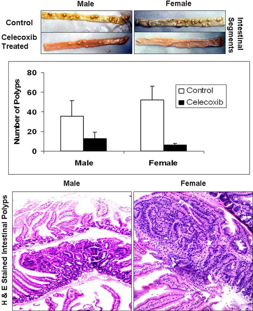
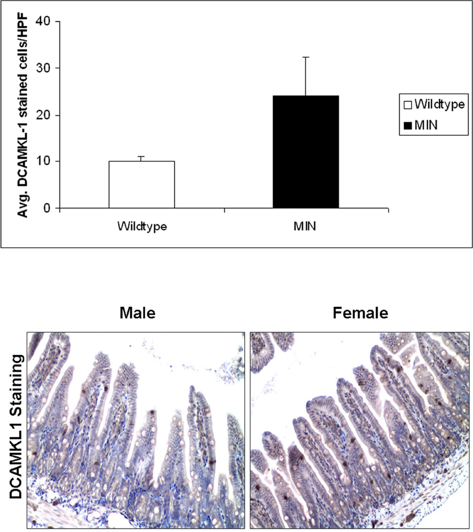
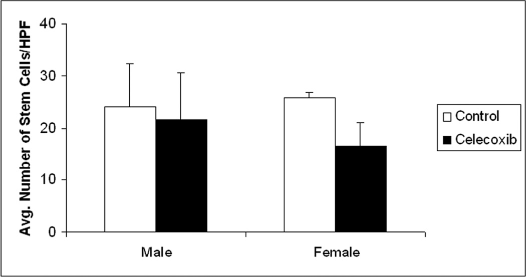
This study was undertaken to examine the gender-sensitivity and chemopreventive responsiveness of celecoxib on intestinal stem-like cells as a biomarker of colon carcino-genesis, using the MIN mouse model. Male and female MIN mice (6-7-weeks old) were randomized to either control diet or to a diet supplemented with celecoxib (1,500 ppm). The animals were euthanized ten weeks later and the intestines were flushed and opened longitudinally to assess tumor count. Small intestinal segments were formalin-fixed and tissue sections were subjected to immunohistochemical evaluation of DCAMKL1, a known marker of stem-like cells. We found that in animals receiving control (AIN 76A diet) alone, female MIN mice had a higher polyp count than males (52.32 ± 13.89 vs. 35.43 ± 16.05; p<0.0005). However, compared to control diet groups, celecoxib supplementation caused a larger reduction in the number of polyps in females than their male cohorts (6.38 ± 1.43 vs. 12.83 ± 6.74; a reduction of 88% in females to 64% in males). Significant differences (p=0.013) were observed in the number of DCAMKL1-stained cells in the crypts of the wild-type (WT) (10.01 ± 1.07 stem cells per high powered field; HPF) compared to the MIN mice (24.15 ± 8.08 stem cells per HPF), illustrating increased stem-like cells in animals that are more prone to neoplasia. DCAMKL1 labeled stem-like cells were equal in number in the male and female groups receiving the control AIN 76A diet alone (females, 25.73 stem-like cells/HPF); males, 24.15 stem-like cells/HPF). However, females showed a greater reduction in the number of DCAMKL1-labeled stem-like cells with celecoxib supplementation than the respective males (16.63 ± 4.23 vs. 21.56 ± 9.06; a reduction of 35.4% in females to 10.7% in males). We conclude that a higher number of stem-like cells in the uninvolved mucosa paralleled tumorigenesis and mirrored greater chemopreventive responsiveness of female MIN mice compared to males.


Figures

Figure 1
Figure 1. Female MIN mice demonstrated higher celecoxib responsiveness to intestinal polyp reduction than males
Total number of tumors in the small intestine (proximal and distal) and the colon for each animal were independently scored by three individuals blinded to the gender group. For easy identification, the tissue samples were cooled on dry-ice as discussed in the “Materials and Methods” section. After calculating the mean of the total number of tumors counted, it was observed that without celecoxib treatment, females developed more polyps than males (52.32±13.89 vs. 35.43±16.05). Celecoxib treated females developed lesser number of tumors than their celecoxib treated males counterparts (6.38±1.43 vs. 12.83±6.74) which reflects that celecoxib treatment resulted in 87.8% decrease (p=2×10⁻¹⁴) in tumor count in females as compared to only 63.7% decrease (p=7×10⁻⁷) in their males counterparts (Figure 1A). Furthermore, the histological analysis of the H&E stained sections of the “Swiss rolls” positively confirmed the adenomatous nature of the intestinal polyps (Figure 1B).
Figure 2
Figure 2. Larger reduction in DCAMKL1 stained intestinal stem-like cells in Female MIN mice in response to chemopreventive celecoxib compared to males
The intestinal tissue sections were subjected to immunohistochemical staining for stem-cell marker DCAMKL1 as described in “Materials and Methods”. The MIN mice (male) on average had 24.15 ± 8.08 stem cells per HPF compared to the wildtype mice which had 10.01 ± 1.07 stem cells per HPF, p=0.013, thus illustrating the increase in stem cells seen in animals more prone to neoplasia (Figure 2A. Furthermore, the number of DCAMKL1 labeled stem-like cells were equal in the male and female groups receiving the control (AIN 76A) diet alone, (females= 25.73 stem cells/HPF, males= 24.15 stem cells/HPF) (Figure 2B). As shown (by intense brown staining), DCAMKL1 had distinct nuclear and cytoplasmic expression pattern. The females that received celecoxib enriched diet however, had a greater reduction in stem-like cells than their male cohorts (16.63±4.23 vs. 21.56±9.06; a reduction of 35.4% in females to 10.7% in males) (Figure 2C).

Similar articles

Cited by

References

    1. Jemal A, Siegel R, Xu J, Ward E. Cancer statistics, 2010. CA Cancer J Clin. 2010;60:277–300. - PubMed
    1. Schoenfeld P, Cash B, Flood A, et al. Colonoscopic screening of average-risk women for colorectal neoplasia. N Engl J Med. 2005;352:2061–2068. - PubMed
    1. Bresalier RS. Chemoprevention of colorectal neoplasia: advances and controversies (the COX-2 story) Curr Opin Gastroenterol. 2007;23:44–47. - PubMed
    1. Guruswamy S, Rao CV. Multi-Target Approaches in Colon Cancer Chemoprevention Based on Systems Biology of Tumor Cell-Signaling. Gene Regul Syst Bio. 2008;2:163–176. - PMC - PubMed
    1. Carpentino JE, Hynes MJ, Appelman HD, Zheng T, Steindler DA, Scott EW, Huang EH. Aldehyde dehydrogenase-expressing colon stem cells contribute to tumorigenesis in the transition from colitis to cancer. Cancer Res. 2009;69:8208–8215. - PMC - PubMed

Publication types

MeSH terms