. 2011 Nov;59(11):1719-31.
doi: 10.1002/glia.21218. Epub 2011 Jul 18.

Motor neuron impairment mediated by a sumoylated fragment of the glial glutamate transporter EAAT2

Affiliations

Motor neuron impairment mediated by a sumoylated fragment of the glial glutamate transporter EAAT2

Emily Foran et al. Glia. 2011 Nov.

Abstract

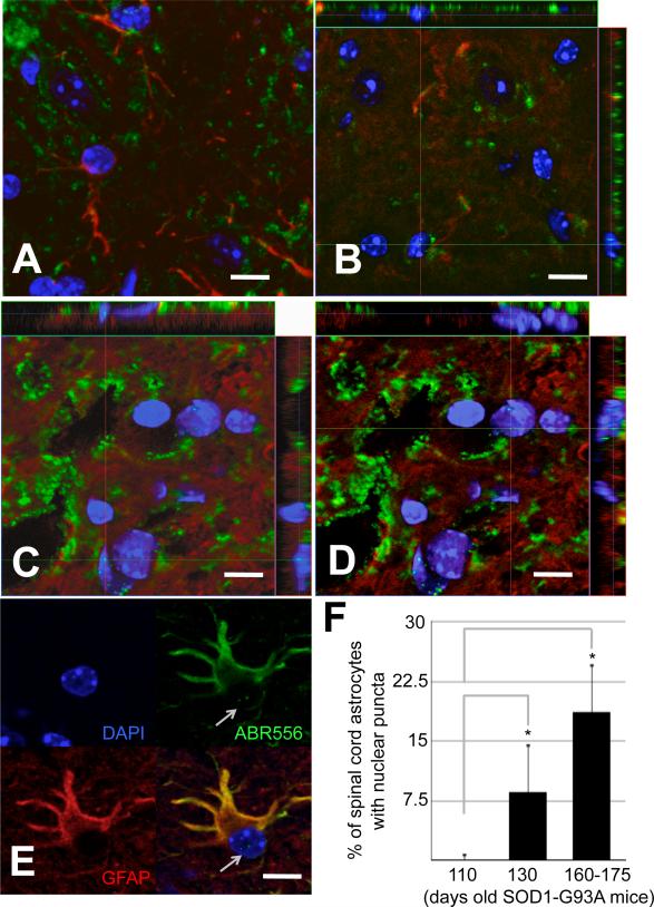
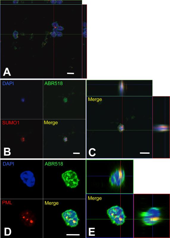
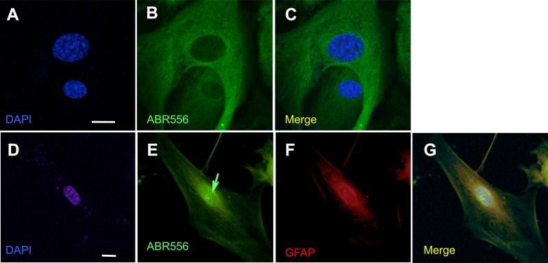
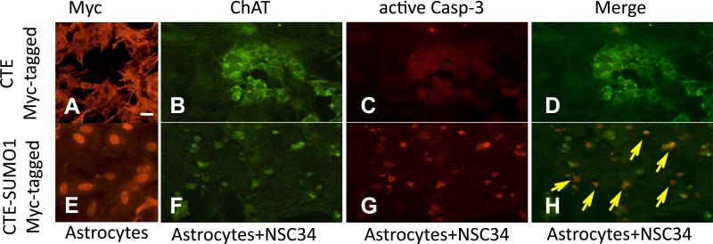
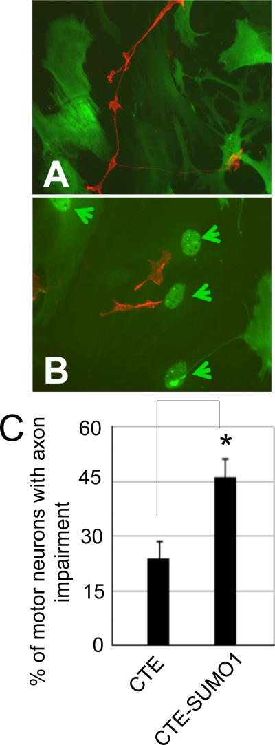
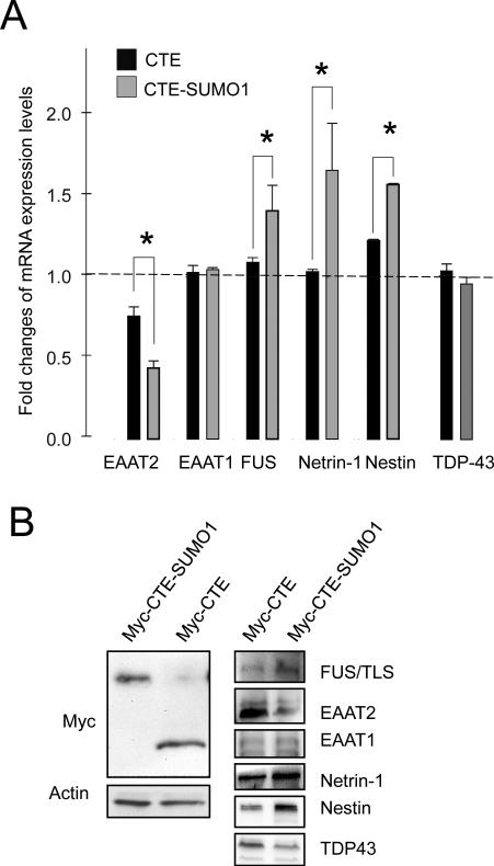
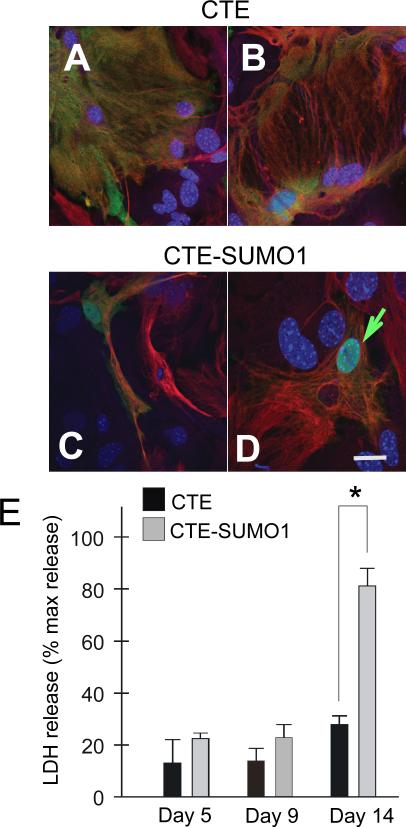
Dysregulation of glutamate handling ensuing downregulation of expression and activity levels of the astroglial glutamate transporter EAAT2 is implicated in excitotoxic degeneration of motor neurons in amyotrophic lateral sclerosis (ALS). We previously reported that EAAT2 (a.k.a. GLT-1) is cleaved by caspase-3 at its cytosolic carboxy-terminus domain. This cleavage results in impaired glutamate transport activity and generates a proteolytic fragment (CTE) that we found to be post-translationally conjugated by SUMO1. We show here that this sumoylated CTE fragment accumulates in the nucleus of spinal cord astrocytes of the SOD1-G93A mouse model of ALS at symptomatic stages of disease. Astrocytic expression of CTE, artificially tagged with SUMO1 (CTE-SUMO1) to mimic the native sumoylated fragment, recapitulates the nuclear accumulation pattern of the endogenous EAAT2-derived proteolytic fragment. Moreover, in a co-culture binary system, expression of CTE-SUMO1 in spinal cord astrocytes initiates extrinsic toxicity by inducing caspase-3 activation in motor neuron-derived NSC-34 cells or axonal growth impairment in primary motor neurons. Interestingly, prolonged nuclear accumulation of CTE-SUMO1 is intrinsically toxic to spinal cord astrocytes, although this gliotoxic effect of CTE-SUMO1 occurs later than the indirect, noncell autonomous toxic effect on motor neurons. As more evidence on the implication of SUMO substrates in neurodegenerative diseases emerges, our observations strongly suggest that the nuclear accumulation in spinal cord astrocytes of a sumoylated proteolytic fragment of the astroglial glutamate transporter EAAT2 could participate to the pathogenesis of ALS and suggest a novel, unconventional role for EAAT2 in motor neuron degeneration.


Figures

Fig.1
Fig.1. EAAT2-immunopositive puncta in spinal cord astrocyte nuclei of SOD1-G93A mice at disease progression
Ten μm spinal cord cross-sections from (A) presymptomatic (70 days old), (B) onset (110 days old) and (C-E) diseased (160-170 days old) G93A-SOD1 mice were immunostained with EAAT2 (ABR556; green; dil. 1:100) and GFAP (red; dil. 1:100) antibodies. The ventral portion of the lumbar spinal cord was imaged. (F) Quantification of GFAP-positive astrocytes displaying C-terminus EAAT2 immunoreactive nuclear punctate. For quantification, spinal cords of 3 animals from each disease stage were cut into 10um cross sections and probed with GFAP, EAAT2 and DAPI. From each mouse the ventral horn of 3 random sections were analyzed with LSM Image software. Asterisk indicates P<0.05 versus onset stage group. Nuclear puncta were absent at presymptomatic stage of disease and not plotted. Scale bar, 10 μm.
Fig.2
Fig.2. The C-terminus fragment of EAAT2 is sumoylated in the nucleus of spinal cord astrocytes of SOD1-G93A mice
Nuclei were isolated from spinal cords of (A) pre-symptomatic (70 days old) and (B-E) diseased G93A-SOD1 mice (~160 days old) and stained for ABR518 (green, 1.5μg/ml), SUMO1 (red, dil. 1:40), and DAPI (blue) (B,C). Puncta immunopositive for the EAAT2 C-terminus antibody ABR518 were also immunoreactive for PML (dil. 1:100) (D,E). No ABR518 immunopositive puncta were detected in nuclei of pre-symptomatic mice (A). Images were taken with 63X oil-immersed objective. Scale bar, 10 μm.
Fig.3
Fig.3. Spinal cord astrocytes cultured from SOD1-G93A diseased mice accumulate a C-terminus fragment of EAAT2 in the nucleus
Astrocytes were isolated from spinal cords of neonatal non-transgenic mice (P2-P4) or from diseased SOD1-G93A mice (~150 days old), expanded in culture, and analyzed by immunofluorescence. Non-transgenic astrocytes did not display nuclear immunoreactivity for EAAT2 (A-C). Instead, there were discrete EAAT2 C-terminus immunopositive puncta (green arrow) in the nucleus of astrocytes isolated from spinal cord of diseased mutant SOD1 mice (D-G), which were not immunoreactive for ABR372 (Suppl.Fig.5A-C). Scale bars, 10 μm.
Fig.4
Fig.4. The C-terminus fragment of EAAT2 is immunoreactive for SUMO1 and partition to PML-nuclear bodies in spinal cord astrocytes cultured from SOD1-G93A diseased mice
Nuclear EAAT2 puncta were also SUMO1 immunoreactive (A-C), although some of them are not (green arrow in D). Puncta either localized in close proximity or were associated with PML immunopositive nuclear bodies (E-H). EAAT2 puncta were immunopositive for both ABR518 and ABR556 (B,F). Scale bar, 10 μm. An higher magnification of panel H is presented in Suppl.Fig.6A.
Fig.5
Fig.5. Nuclear accumulation in astrocytes of CTE-SUMO1 leads to activation of caspase-3 in co-cultured NSC-34 cells
Nontransgenic spinal cord astrocytes were transduced with either AdV-myc-CTE or with AdV-myc-CTE-SUMO1 (MOI 2). Three days post transduction NSC34 cells were plated on the bed of either (A) Myc-CTE or (E) Myc-CTE-SUMO1 expressing astrocytes. Nuclear localization of CTE-SUMO1 is clearly noticeable (E) compared to the cytosolic distribution of CTE (A). NSC34 cells can be identified by ChAT (dil.1:100) staining (B,F). NSC34 cells plated on CTE-SUMO1 astrocytes were positive for active caspase 3 immunostaining (dil. 1:100; arrows in merged panel) (G,H) compared to NSC34 cells plated on CTE-expressing astrocytes (C,D). Representative fluorescence microscopy images are displayed. Scale bar, 10 μm.
Fig.6
Fig.6. Spinal cord astrocytes displaying nuclear localization of CTE-SUMO1 are toxic to motor neurons in culture
Nontransgenic spinal cord astrocytes were transfected with either myc-CTE or with myc-CTE-SUMO1 plasmid cDNA. After six days purified non-transgenic motor neurons were plated on either Myc-CTE (A) or Myc-CTE-SUMO1 expressing (B) astrocytes. Expression of the constructs was assessed with anti-Myc antibody (dil. 1:100; green). Motor neurons plated on spinal cord astrocytes displaying nuclear accumulation of CTE-SUMO1 (arrowheads) had a larger percent of cells with axonal impairment (C). Motor neurons were immunostained with P75NTR antibody (red; dil. 1:100). Representative images of 4 different experiments are presented. *P<0.01, one-tail t-test.
Fig.7
Fig.7. Validation of microarray data
Nontransgenic spinal cord astrocytes were transfected with Myc-CTE or Myc-CTE-SUMO1. Analysis was performed either by qRT-PCR or western blot. (A) Six genes from the microarray experiment, which displayed statistically significant changes in the CTE-SUMO1 group, were tested with quantitative real time PCR. Data are normalized for β-actin mRNA and are the average of 6 independent experiments. Of these genes, EAAT1 and TDP-43 did not show significant differences in RNA expression levels. Two-tail t-test, *P<0.05. (B) Protein expression levels of the same six genes were also tested by western blot to confirm that the mRNA changes carried on to protein changes. TDP-43 protein expression levels decreased in contrast with the qPCR data set but in agreement with microarray analysis. Spinal cord astrocytes showed equal levels of CTE or CTE-SUMO1 assessed by western blot with anti-Myc antibody. A representative western blot analysis is shown. Astrocytes expressing CTE or CTE-SUMO1 were lysed in modified RIPA buffer and run on a 16% TrisTricine gel, transferred to PVDF membrane and probed as indicated.
Fig.8
Fig.8. Persistent nuclear accumulation of CTE-SUMO1 is intrinsically toxic to astrocytes
Nontransgenic spinal cord astrocytes were transfected with either myc-CTE or myc-CTE-SUMO1. Lactate dehydrogenase (LDH) levels released in the medium was assessed at days 5, 9, and 14 post-transfection using the Clontech LDH cytotoxicity detection kit (cat.# 630117) following manufacturer's instructions and compared to LDH released by exposure of the cells to 30 min of 1% Triton X-100. At day 14 a subset of transfected astrocytes were fixed and stained with GFAP (red), Myc (green) and DAPI. Astrocytes expressing CTE retained their normal morphology after two weeks in culture (A), while nuclear accumulation of CTE-SUMO1 in led to dramatic altered morphology at 14 days in vitro in astrocytes including elongated processes (C) and large vacuoles (D). In A-D are shown representative confocal microscopy images. Results in E are the average ± s.e.m. of 3 independent experiments, each run in triplicate.

Similar articles

Cited by

References

    1. Acarin L, Villapol S, Faiz M, Rohn TT, Castellano B, Gonzalez B. Caspase-3 activation in astrocytes following postnatal excitotoxic damage correlates with cytoskeletal remodeling but not with cell death or proliferation. Glia. 2007;55(9):954–65. - PubMed
    1. Bernardi R, Pandolfi PP. Structure, dynamics and functions of promyelocytic leukaemia nuclear bodies. Nat Rev Mol Cell Biol. 2007;8(12):1006–16. - PubMed
    1. Blackburn D, Sargsyan S, Monk PN, Shaw PJ. Astrocyte function and role in motor neuron disease: a future therapeutic target? Glia. 2009;57(12):1251–64. - PubMed
    1. Boston-Howes W, Gibb SL, Williams EO, Pasinelli P, Brown RH, Jr., Trotti D. Caspase-3 cleaves and inactivates the glutamate transporter EAAT2. J Biol Chem. 2006;281(20):14076–84. - PubMed
    1. Brenner M, Kisseberth WC, Su Y, Besnard F, Messing A. GFAP promoter directs astrocyte-specific expression in transgenic mice. J Neurosci. 1994;14(3 Pt 1):1030–7. - PMC - PubMed

Publication types

MeSH terms