

A whole brain fMRI atlas generated via spatially constrained spectral clustering
- PMID: 21769991
- PMCID: PMC3838923
- DOI: 10.1002/hbm.21333
A whole brain fMRI atlas generated via spatially constrained spectral clustering
Abstract
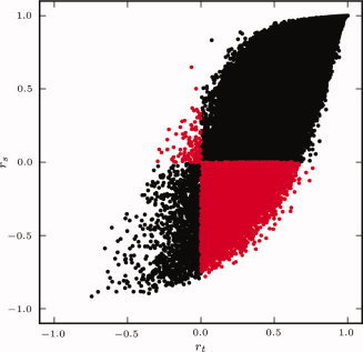
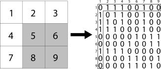
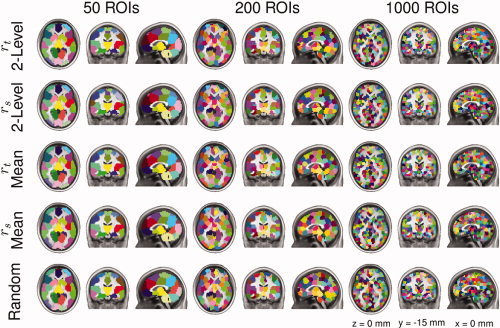
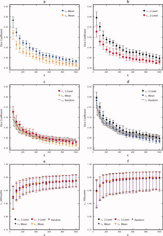
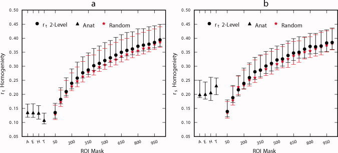
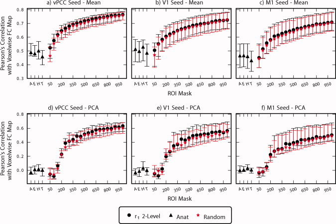
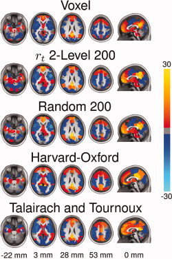
Connectivity analyses and computational modeling of human brain function from fMRI data frequently require the specification of regions of interests (ROIs). Several analyses have relied on atlases derived from anatomical or cyto-architectonic boundaries to specify these ROIs, yet the suitability of atlases for resting state functional connectivity (FC) studies has yet to be established. This article introduces a data-driven method for generating an ROI atlas by parcellating whole brain resting-state fMRI data into spatially coherent regions of homogeneous FC. Several clustering statistics are used to compare methodological trade-offs as well as determine an adequate number of clusters. Additionally, we evaluate the suitability of the parcellation atlas against four ROI atlases (Talairach and Tournoux, Harvard-Oxford, Eickoff-Zilles, and Automatic Anatomical Labeling) and a random parcellation approach. The evaluated anatomical atlases exhibit poor ROI homogeneity and do not accurately reproduce FC patterns present at the voxel scale. In general, the proposed functional and random parcellations perform equivalently for most of the metrics evaluated. ROI size and hence the number of ROIs in a parcellation had the greatest impact on their suitability for FC analysis. With 200 or fewer ROIs, the resulting parcellations consist of ROIs with anatomic homology, and thus offer increased interpretability. Parcellation results containing higher numbers of ROIs (600 or 1,000) most accurately represent FC patterns present at the voxel scale and are preferable when interpretability can be sacrificed for accuracy. The resulting atlases and clustering software have been made publicly available at: http://www.nitrc.org/projects/cluster_roi/.
Copyright © 2011 Wiley Periodicals, Inc.
Figures
References
-
- Ashburner J, Friston KJ ( 2005): Unified segmentation. Neuroimage 26: 839–851. - PubMed
-
- Baumgartner R, Scarth G, Teichtmeister C, Somorjai R, Moser E ( 1997): Fuzzy clustering of gradient‐echo functional MRI in the human visual cortex. Part I: reproducibility. J Magn Reson Imaging 7: 1094–1101. - PubMed
-
- Bellec P, Perlbarg V, Jbabdi S, Pelegrini‐Issac M, Anton JL, Doyon J, Benali H ( 2006): Identification of large‐scale networks in the brain using fMRI. Neuroimage 29: 1231–1243. - PubMed
-
- Biswal BB, Mennes M, Zuo XN, Gohel S, Kelly C, Smith SM, Beckmann CF, Adelstein JS, Buckner RL, Colcombe S, Dogonowski AM, Ernst M, Fair D, Hampson M, Hoptman MJ, Hyde JS, Kiviniemi VJ, Kötter R, Li SJ, Lin CP, Lowe MJ, Mackay C, Madden DJ, Madsen KH, Margulies DS, Mayberg HS, McMahon K, Monk CS, Mostofsky SH, Nagel BJ, Pekar JJ, Peltier SJ, Petersen SE, Riedl V, Rombouts SA, Rypma B, Schlaggar BL, Schmidt S, Seidler RD, Siegle GJ, Sorg C, Teng GJ, Veijola J, Villringer A, Walter M, Wang L, Weng XC, Whitfield‐Gabrieli S, Williamson P, Windischberger C, Zang YF, Zhang HY, Castellanos FX, Milham MP ( 2010): Toward discovery science of human brain function. Proc Natl Acad Sci U S A 107: 4734–4739. - PMC - PubMed
Publication types
MeSH terms
Grants and funding
LinkOut - more resources
Full Text Sources
Other Literature Sources
