. 2011 Aug 23;50(33):7117-31.
doi: 10.1021/bi200905x. Epub 2011 Jul 26.

Efficient isolation of Pseudomonas aeruginosa type III secretion translocators and assembly of heteromeric transmembrane pores in model membranes

Affiliations

Efficient isolation of Pseudomonas aeruginosa type III secretion translocators and assembly of heteromeric transmembrane pores in model membranes

Fabian B Romano et al. Biochemistry. .

Abstract

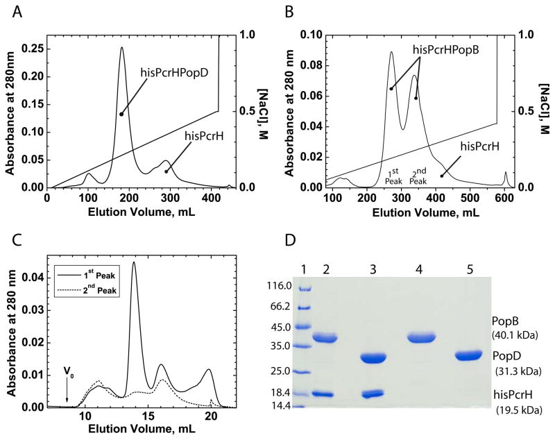
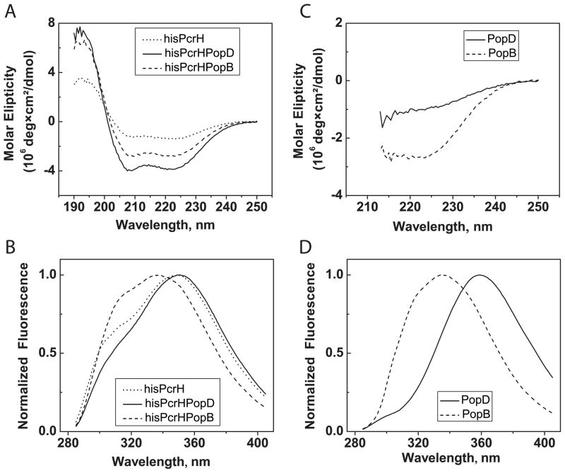
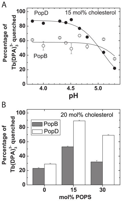
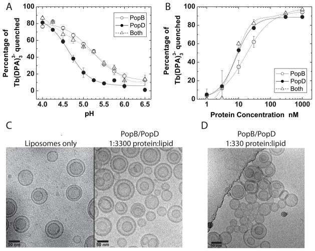
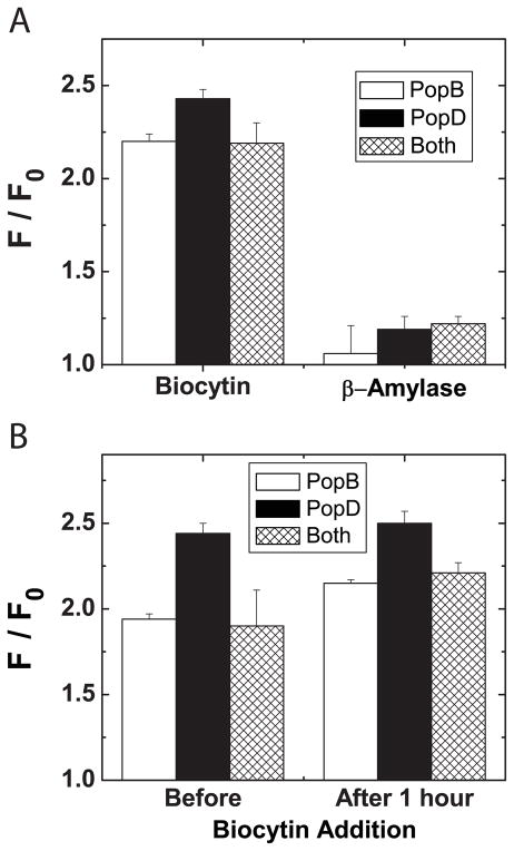
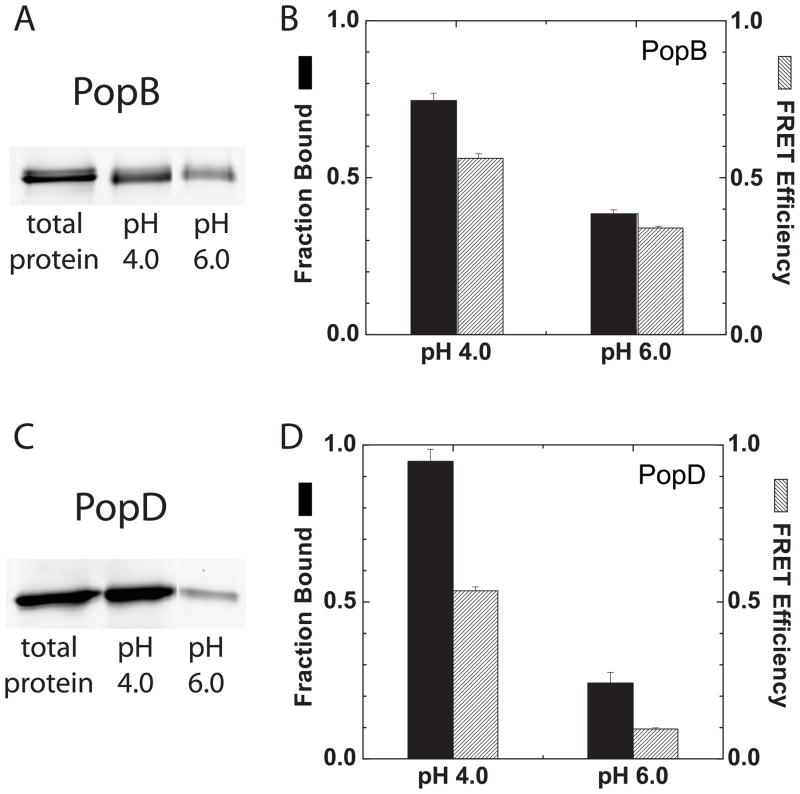
Translocation of bacterial toxins or effectors into host cells using the type III secretion (T3S) system is a conserved mechanism shared by many Gram-negative pathogens. Pseudomonas aeruginosa injects different proteins across the plasma membrane of target cells, altering the normal metabolism of the host. Protein translocation presumably occurs through a proteinaceous transmembrane pore formed by two T3S secreted protein translocators, PopB and PopD. Unfolded translocators are secreted through the T3S needle prior to insertion into the target membrane. Purified PopB and PopD form pores in model membranes. However, their tendency to form heterogeneous aggregates in solution had hampered the analysis of how these proteins undergo the transition from a denatured state to a membrane-inserted state. Translocators were purified as stable complexes with the cognate chaperone PcrH and isolated from the chaperone using 6 M urea. We report here the assembly of stable transmembrane pores by dilution of urea-denatured translocators in the presence of membranes. PopB and PopD spontaneously bound liposomes containing anionic phospholipids and cholesterol in a pH-dependent manner as observed by two independent assays, time-resolved Förster resonance energy transfer and sucrose-step gradient ultracentrifugation. Using Bodipy-labeled proteins, we found that PopB interacts with PopD on the membrane surface as determined by excitation energy migration and fluorescence quenching. Stable transmembrane pores are more efficiently assembled at pH <5.0, suggesting that acidic residues might be involved in the initial membrane binding and/or insertion. Altogether, the experimental setup described here represents an efficient method for the reconstitution and analysis of membrane-inserted translocators.


Figures

Figure 1
Figure 1. Purification of the hisPcrH-translocator complexes
(A) The fractions containing the hisPcrH-PopD complex isolated after the first IMAC purification step were dialyzed and loaded into a Q-Sepharose AEC column and eluted using a linear NaCl gradient. The peaks containing hisPcrH and hisPcrH-PopD are indicated. (B) The hisPcrH-PopB complex was purified as described for hisPcrH-PopD. The first two large peaks that eluted from the AEC contained the hisPcrH-PopB complexes and the shoulder that eluted around 400 mL contained hisPcrH. (C) SEC analysis of aliquots corresponding to the 1st Peak and the 2nd Peak illustrated in B. (D) SDS-PAGE analysis of purified proteins. Lane 1, molecular weight markers; lane 2, hisPcrH-PopB; lane 3, hisPcrH-PopD; lanes 4 and 5, urea-isolated PopB and PopD, respectively.
Figure 2
Figure 2. Characterization of purified translocators
(A) Far UV CD spectra of the SEC-isolated hisPcrH, hisPcrH-PopD, and hisPcrH-PopB recorded in buffer E, total protein concentration was 3.0 μM. (B) Normalized fluorescence emission spectra of hisPcrH, hisPcrH-PopD, and hisPcrH-PopB recorded in buffer E. Excitation wavelength was 278 nm, total protein concentration was 2.4 μM. (C) Far UV CD spectra of purified PopD and PopB recorded in phosphate buffer 20 mM, pH 7.5 supplemented with urea 6 M, protein concentration was 2.4 μM. (D) Normalized fluorescence emission spectra of PopD and PopB in phosphate buffer 20 mM, pH 7.5 supplemented with urea 6 M. Excitation wavelength was 278 nm, total protein concentration was 2.4 μM.
Figure 3
Figure 3. Effect of pH and lipid composition on the pore forming activity of the urea-isolated translocators
Pore formation was determined as the percentage of the encapsulated Tb(DPA)33- that was quenched by EDTA as detailed in experimental procedures. (A) Urea-isolated PopB or PopD were directly diluted into a solution of 50 mM sodium acetate buffered at the indicated pH, containing membranes. The pore forming activity of the urea-isolated translocators increased at acidic pH. The total lipid concentration was 0.1 mM and the liposomes composition was 35 mol % POPC, 18 mol % POPS, 24 mol % POPE, 17 mol% SM, and 15 mol % cholesterol. Protein:lipid ratio was 1:1000. (B) Effect of POPS on the pore formation activity of urea-isolated translocators. The activity was measured as described in A, the pH was buffered at 4.3, and the lipid composition was POPC, 20 mol % cholesterol, and the indicated concentration of POPS.
Figure 4
Figure 4. Pore forming activity of the urea-isolated translocators
Pore formation was determined as the percentage of the encapsulated Tb(DPA)33- that was quenched by EDTA as detailed in experimental procedures. (A) Urea-isolated PopB, PopD, or an equimolar mixture of both proteins were diluted into a buffer solution containing membranes at the indicated pH. Total concentration of lipids was 0.15 mM. Total protein concentration was 60 nM for the PopB and PopD traces (protein:lipid ratio 1/2500), and 120 nM when PopB and PopD where added together (protein:lipid ratio 1/1250). (B) Concentration-dependent pore formation by PopD, PopB (protein:lipid ratio ranged from 1/105 to 1/100), or an equimolar mixture of both proteins diluted into buffer D containing 0.1 mM total lipids. When both proteins were added together the sample contained twice as much total protein than the concentration indicated in the graph (protein:lipid ratio ranged from 1/5.104 to 1/50). (C) Cryo-electron micrographs of liposomes with and without PopB/PopD at the indicated protein:lipid ratios. Buffer B was added to the control sample with only liposomes to assess any effect in membrane structure due to residual urea concentration. Lipid concentration was 5 mM in all cases and total protein concentration was 1.5 μM or 15 μM. The POPC:cholesterol:POPS molar ratio was 65:20:15 in all panels.
Figure 5
Figure 5. PopD and PopB form discrete and stable pores in model membranes
(A) The passage of biocytin (~10Å diameter), biotin-labeled β-amylase (~50Å diameter), or streptavidin-Bodipy (~50Å diameter) through the pores formed by PopD, PopB, or an equimolar mixture of the translocators was measured as detailed in experimental procedures. Total lipid concentration was 100 μM, and protein concentration of the proteins was 100 nM (protein:lipid ratio was 1/1000 for individual proteins and 1/500 when added together). The fluorescence intensity of encapsulated streptavidin-Bodipy was measured before (F0) or after the incubation for 60 min at 25°C with the translocators (F). Only biocytin was able to diffuse through the formed pores. (B) The stability of the formed pores was examined by measuring the increase on the fluorescence intensity of encapsulated streptavidin-Bodipy when biocytin was present in the external buffer solution before the addition of the translocators, or added after 1 hr of incubation with the translocators. The discrete pores formed by PopB, PopD, and an equimolar mixture of the proteins remained open after1hr of incubation.
Figure 6
Figure 6. Acidic pH enhances translocator membrane binding
(A) PopBS164C-Bodipy membrane binding was measured using the liposome floatation assay described in experimental procedures. PopBS164C-Bodipy (0.4 μM) was incubated with membranes (2 mM total lipids) for 1 hr at 20–23°C. Typical SDS-PAGE analysis is indicated showing the amount of total protein added, and the amount of bound protein isolated after incubation with membranes at the indicated pH. Protein:lipid ratio was 1:5000. Gels were scanned for Bodipy fluorescence using a phosphoimager. (B) Quantification of PopBS164C-Bodipy binding measured by liposome floatation assay (Fraction Bound, black bars). Data was normalized against the total amount of protein used in the binding reaction. Each bar represents the average and range of at least two independent experiments. FRET efficiency (grey bars) for PopBS164C-Bodipy binding determined by time-resolved FRET as described in experimental procedures. PopBS164C-Bodipy (120 nM) was incubated with membranes (total lipid concentration 0.3 mM) as described in A) (protein:lipid ratio was 1:2500). Each bar represents the average and range of two independent experiments. (C) and (D) show the analysis for PopDF223C-Bodipy binding as described in A) and B) for PopBS164C-Bodipy. The POPC:cholesterol:POPS molar ratio was 65:20:15 in all panels.

References

    1. Hueck CJ. Type III protein secretion systems in bacterial pathogens of animals and plants. Microbiol Mol Biol Rev. 1998;62:379–433. - PMC - PubMed
    1. Galan JE, Wolf-Watz H. Protein delivery into eukaryotic cells by type III secretion machines. Nature. 2006;444:567–573. - PubMed
    1. Cornelis GR. The type III secretion injectisome. Nat Rev Micro. 2006;4:811–825. - PubMed
    1. Frank D. The exoenzyme S regulon of Pseudomonas aeruginosa. Mol Microbiol. 1997;26:621–629. - PubMed
    1. Hauser AR, Cobb E, Bodi M, Mariscal D, Valles J, Engel JN, Rello J. Type III protein secretion is associated with poor clinical outcomes in patients with ventilator-associated pneumonia caused by Pseudomonas aeruginosa. Crit Care Med. 2002;30:521–528. - PubMed

Publication types

MeSH terms

LinkOut - more resources