. 2011 Sep;28(9):1827-43.
doi: 10.1089/neu.2011.1911. Epub 2011 Sep 8.

The effect of progesterone dose on gene expression after traumatic brain injury

Affiliations

The effect of progesterone dose on gene expression after traumatic brain injury

Gail D Anderson et al. J Neurotrauma. 2011 Sep.

Abstract

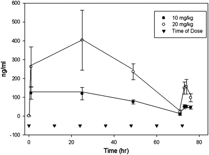
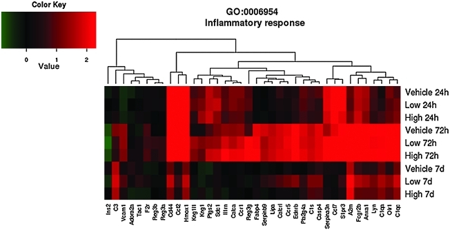
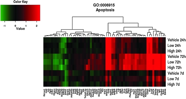
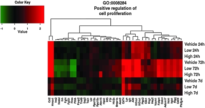
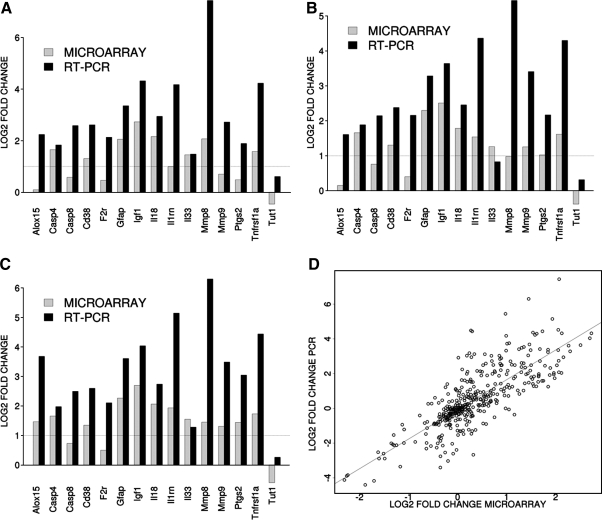
Microarray-based transcriptional profiling was used to determine the effect of progesterone in the cortical contusion (CCI) model. Gene ontology (GO) analysis then evaluated the effect of dose on relevant biological pathways. Treatment (vehicle, progesterone 10 mg/kg or 20 mg/kg given i.p.) was started 4 h post-injury and administered every 12 h post-injury for up to 72 h, with the last injection 12 hr prior to death for the 24 h and 72 h groups. In the CCI-injured vehicle group compared to non-injured animals, expression of 1,114, 4,229, and 291 distinct genes changed >1.5-fold (p<0.05) at 24 h, 72 h, and 7 days, respectively. At 24 h, the effect of low-dose progesterone on differentially expressed genes was <20% the effect of higher dose compared to vehicle. GO analysis identified a significant effect of low- and high-dose progesterone treatment compared to vehicle on DNA damage response. At 72 h, high-dose progesterone treatment compared to vehicle affected expression of almost twice as many genes as did low-dose progesterone. Both low- and high-dose progesterone resulted in expression of genes regulating inflammatory response and apoptosis. At 7 days, there was only a modest difference in high-dose progesterone compared to vehicle, with only 14 differentially expressed genes. In contrast, low-dose progesterone resulted in 551 differentially expressed genes compared to vehicle. GO analysis identified genes for the low-dose treatment involved in positive regulation of cell proliferation, innate immune response, positive regulation of anti-apoptosis, and blood vessel remodeling.


Figures

FIG. 1.
FIG. 1.
Histology plate. Shown in the top panel are representative dorsal images depicting normal (grade 2) levels of damage at various time points post-CCI. The bottom panel shows a cortical slab before and after tissue punching to remove the core of the injury for molecular analysis.
FIG. 2.
FIG. 2.
Mean plasma concentrations (and standard deviations) of progesterone following administration of 10 mg/kg or 20 mg/kg i.p. at time 0, 12 h, 24 h, 36h, 48 h, 60 h, and 72 h.
FIG. 3.
FIG. 3.
Effects of traumatic brain injury and progesterone treatment on expression of genes involved in inflammatory response. Genes belonging to the inflammatory response gene ontology category (GO:0006954) whose expression changed more than 1.5-fold (up or down, p<0.05) in at least one of the three comparisons “Low progesterone treatment versus vehicle treatment”, “High progesterone treatment versus vehicle treatment”, or “Low progesterone treatment versus high progesterone treatment,” are displayed. The rows show the average expression of the vehicle, and low and high progesterone experimental groups at 24 h, 72 h, and 7 days. Each column shows the expression of a single gene. Red indicates that expression was higher compared to the average of the uninjured sham group, green indicates the opposite, and black means no change. Abbreviations: A2m, alpha-2-macroglobulin; Adora2a, adenosine A2a receptor; Anxa1, annexin A1; C1qa, complement component 1, q subcomponent, alpha polypeptide; C1qc,complement component 1, q subcomponent C chain. group IVA (cytosolic, calcium-dependent); C1s, complement component 1, s subcomponent; C3, complement component 3; Calca, calcitonin/calcitonin-related polypeptide, alpha; Calcr l, calcitonin recptor-like; Casp4, caspase 4, apoptosis-related cystine peptidase; Ccl12, chemokine (C-C motif) ligand 12; Ccl17, chemokine (C-C motif) ligand17; Ccr1, chemokine (C-C) receptor 1; Ccr5, chemokine (C-C motif) receptor 5; Cd44, Cd44 molecule; Ednrb, endothelin receptor type b; F2r, coagulation factor II (thrombin) receptor; Fabp4, fatty acid binding protein 4, adipocyte; Fcgr2b, FC fragment of IgG, low affinity llb, receptor (CD32); Hmox1, heme oxygenase (decycling) 1; Il1rn, interleukin 1 receptor antagonist; Ins2, insulin 2; Kng1l1, kininogen 1-like 1; Kng1, kininogen 1; Lipa, lipase A, lysomsomal acid, cholesterol esterase; Lyn, v-yes-1 Yamagtuchi sarcoma viral related oncogene homolog; Olr1, oxidized low density lipoprotein (lectin-like) receptor 1; Pla2g4a, phospholipase A2, group IVA (cytosolic, calcium-dependent); Ptgs2, prostaglandin-endoperoxide synthase 2; Reg3a, regenerating islet-derived 3 alpha; Reg3b, regenerating islet-derived 3 beta; Reg3g, regenerating islet-derived 3 gamma; S1pr3, serine (or cystine) peptidase inhibitor, clade A, member 3N; Sdc1, syndecan1; Tac1, tachykinin 1.
FIG. 4.
FIG. 4.
Effects of traumatic brain injury and progesterone treatment on expression of genes involved in apoptosis. Genes belonging to the apoptosis gene ontology category (GO: 0006915) whose expression changed more than 1.5-fold (up or down, p<0.05) in at least one of the three comparisons “Low progesterone treatment versus vehicle treatment”, “High progesterone treatment versus vehicle treatment”, or “Low progesterone treatment versus high progesterone treatment,” are displayed. The rows show the average expression of the vehicle and low and high progesterone experimental groups at 24 h, 72 h, and 7 days. Each column shows the expression of a single gene. Red indicates that expression was higher compared to the average of the uninjured sham group, green indicates the opposite, and black means no change. Abbreviations: Adam17, ADAM metallopeptidase domain 17; Adora2a, adenosine A2a receptor; Agtr1a, angiotensin II receptor, type 1; Aldh1a1, aldehyde dehydrogenase 1 family, member A1; Alox15, arachidonate 15-lipoxygenase; Anxa1, annexin A1; Aph1b, anterior pharynx defective 1 homolog B (C. elegans); Api5, apoptosis inhibitor 5; Bag4, BCL2-associated athanogene 4; Brca2, breast cancer 2, early onset; Bub1, budding uninhibited by benzimidazoles 1 homolog (S. cerevisiae); C7, complement component 7; Casp4, caspase 4, apoptosis-related cysteine peptidase; Ccl2, chemokine (C-C motif) ligand 2; Cd44, CD44 molecule (Indian blood group); Cd74, CD74 molecule, major histocompatibility complex, class II invariant chain; Cflar, CASP8 and FADD-like apoptosis regulator; Ctrb1, chymotrypsinogen B1; Dad1, defender against cell death 1; Dram2, transmembrane protein 77; Dusp1, dual specificity phosphatase 1; Ednrb, endothelin receptor type B; Egln3, egl nine homolog 3 (C. elegans); F2r, coagulation factor II (thrombin) receptor; Fastkd2, FAST kinase domains 2; Fgf4, fibroblast growth factor 4; Gfra1, GDNF family receptor alpha 1; Hells, helicase, lymphoid-specific; Hipk1, homeodomain interacting protein kinase 1; Hmox1, heme oxygenase (decycling) 1; Hras, v-Ha-ras Harvey rat sarcoma viral oncogene homolog; Hrk, harakiri, BCL2 interacting protein (contains only BH3 domain); Hspb1, heat shock 27kDa protein 1, Igf1, insulin-like growth factor 1 (somatomedin C); Ihh, Indian hedgehog; Il1rn, interleukin 1 receptor antagonist; Il7r, interleukin 7 receptor; Ins2, insulin 2; Lcn2, lipocalin 2; Ldha, lactate dehydrogenase A; Lifr, leukemia inhibitory factor receptor alpha; Mmp9, matrix metallopeptidase 9; Mst4, serine/threonine protein kinase MST4; Nqo1, NAD(P)H dehydrogenase, quinone 1; Nr4a2, nuclear receptor subfamily 4, group A, member 2; Nrbp2, nuclear receptor binding protein 2; Obscn, obscurin, cytoskeletal calmodulin and titin-interacting RhoGEF; Pla2g4a, phospholipase A2, group IVA (cytosolic, calcium-dependent); Ptgs2, prostaglandin-endoperoxide synthase 2 (prostaglandin G/H synthase and cyclooxygenase); Rab27a, RAB27A, member RAS oncogene family; Rasgrf2, Ras protein-specific guanine nucleotide-releasing factor 2; Rb1cc1, RB1-inducible coiled-coil 1; Rbpj, recombination signal binding protein for immunoglobulin kappa J region; Scg2, secretogranin II (chromogranin C); Serpinb9, serpin peptidase inhibitor, clade B (ovalbumin), member 9; Sh2d1b1, SH2 domain containing 1B; Sfrp1, secreted frizzled-related protein 1; Stk17b, serine/threonine kinase 17b; Tnfrsf11b, tumor necrosis factor receptor superfamily, member 11b; Top2a, topoisomerase (DNA) II alpha 170kDa; Traf6, TNF receptor-associated factor 6; Ube2v2, ubiquitin-conjugating enzyme E2 variant 2.
FIG. 5.
FIG. 5.
Effects of traumatic brain injury and progesterone treatment on expression of genes involved in positive regulation of cell proliferation. Genes belonging to the positive regulation of cell proliferation gene ontology category (GO: 0008284) whose expression changed more than 1.5-fold (up or down, p<0.05) in at least one of the three comparisons “Low progesterone treatment versus vehicle treatment”, “High progesterone treatment versus vehicle treatment”, and “Low progesterone treatment versus high progesterone treatment” are displayed. The rows show the average expression of the vehicle and low and high progesterone experimental groups at 24 h, 72 h, and 7 days. Each column shows the expression of a single gene. Red indicates that expression was higher compared to the average of the uninjured sham group, green indicates the opposite, and black means no change. Abbreviations: Adam17, ADAM metallopeptidase domain 17; Alox15, arachidonate 15-lipoxygenase; Bmpr1a, bone morphogenetic protein receptor, type 1A; Calcrl, calcitonin receptor-like; Ccl12, chemokine (C-C motif) ligand 12; Cd38, Cd38 molecule; F2r, coagulation factor (thrombin) receptor; Fabp4, fatty acid binding protein 4, adipocyte; Fgfr4, fibroblast growth factor receptor 4; Foxp2, forkhead box P2; Hey2, hairy/enhancer of split related to YRPW motif 2; Hipk1, homeodomain interacting protein kinase 1; Hras, Harvey rat sarcoma virus oncogene; Htr2a, 5-hydroxytryptamine (serotonin) receptor 2A; igsf1, immunoglobulin superfamily, member 1; ihh, Indian hedgehog; il13ra1, interleukin 13 receptor, alpha 1; Ins2, insulin 2; itgb1, integrin beta 1 (fibronectin receptor beta); Klf5, Kruppel-like factor 5; Lifr, leukemia inhibitory factor receptor alpha; Lyn, v-yes-1 Yamaguchi sarcoma viral related oncogene homolog; Nck1, NCK adaptor protein 1; Nodal, nodal homolog (mouse); Odc1, ornithine decarboxylase 1; Ptgs2, prostaglandin-endoperoxide synthase 2; Pla2g4a, phospholipase A2, Pthlh, parathyroid hormone-like hormone; Rbpj, recombination signal binding protein for immunoglobulin kappa J region; Rps15a, ribosomal protein S15a; Stat1, signal transducer and activator of transcription 1; Scg2; secetogranin II (chromogranin c); Tac1, tachykinin 1; Traf6, Tnf receptor-associated factor 6; Vcam1, vascular cell adhesion molecule 1.
FIG. 6.
FIG. 6.
TaqMan-based RT-PCR validation of the microarray data for the selected genes: Casp 4, caspase 4; Casp 8, caspase 8; Cd 38, CD38 molecule, F2r, coagulation factor II (thrombin) receptor; Gfap, glial fibrillary acidic protein; Igf1, insulin-like growth factor 1; Il18, interleukin 18; Il1rn, interleukin 1 receptor antagonist; Il33, interleukin 33; Mmp8, matrix metallopeptidase 8; Mmp9, matrix metallopeptidase 9; Ptgs2, prostaglandin-endoperoxide synthase 2; Tnfrsf1a, tumor necrosis factor receptor superfamily, member 1a; Tut1; terminal uridylyl transferase 1. The RT-PCR data were normalized to the housekeeping gene GAPDH. In order to compare the microarray data to the RT-PCR data, the microarray data for each gene was divided by the GAPDH data as it was measured by the microarray analysis. A–C: The gray bars show the microarray data and the black bars display the RT-PCR data for the contrast. Panels A, B and C show the data from the 72-h time point for the contrasts vehicle versus sham, low prostaglandin versus sham and high prostaglandin versus sham, respectively. Panel D shows the overall correlation between the microarray data and the RT-PCR for all contrasts.

References

    1. Allison D.B. Cui X. Page G.P. Sabripour M. Microarray data analysis: from disarray to consolidation and consensus. Nat. Rev. Genet. 2006;7:55–65. - PubMed
    1. Aragones G. Ferre R. Lazaro I. Cabre A. Plana N. Merino J. Heras M. Girona J. Masana L. Fatty acid-binding protein 4 is associated with endothelial dysfunction in patients with type 2 diabetes. Atherosclerosis. 2010;213:329–331. - PubMed
    1. Bazan N.G. Rodriguez de Turco E.B. Allan G. Mediators of injury in neurotrauma: intracellular signal transduction and gene expression. J. Neurotrauma. 1995;12:791–814. - PubMed
    1. Belelli D. Casula A. Ling A. Lambert J.J. The influence of subunit composition on the interaction of neurosteroids with GABA(A) receptors. Neuropharmacology. 2002;43:651–661. - PubMed
    1. Brain S.D. Williams T.J. Tippins J.R. Morris H.R. MacIntyre I. Calcitonin gene-related peptide is a potent vasodilator. Nature. 1985;313:54–56. - PubMed

Publication types

LinkOut - more resources