Lophotrochozoan neuroanatomy: An analysis of the brain and nervous system of Lineus viridis(Nemertea) using different staining techniques
- PMID: 21771310
- PMCID: PMC3160363
- DOI: 10.1186/1742-9994-8-17
Lophotrochozoan neuroanatomy: An analysis of the brain and nervous system of Lineus viridis(Nemertea) using different staining techniques
Abstract
Background: The now thriving field of neurophylogeny that links the morphology of the nervous system to early evolutionary events relies heavily on detailed descriptions of the neuronal architecture of taxa under scrutiny. While recent accounts on the nervous system of a number of animal clades such as arthropods, annelids, and molluscs are abundant, in depth studies of the neuroanatomy of nemerteans are still wanting. In this study, we used different staining techniques and confocal laser scanning microscopy to reveal the architecture of the nervous system of Lineus viridis with high anatomical resolution.
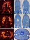
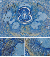
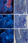
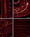
Results: In L. viridis, the peripheral nervous system comprises four distinct but interconnected nerve plexus. The central nervous system consists of a pair of medullary cords and a brain. The brain surrounds the proboscis and is subdivided into four voluminous lobes and a ring of commissural tracts. The brain is well developed and contains thousands of neurons. It does not reveal compartmentalized neuropils found in other animal groups with elaborate cerebral ganglia.
Conclusions: The detailed analysis of the nemertean nervous system presented in this study does not support any hypothesis on the phylogenetic position of Nemertea within Lophotrochozoa. Neuroanatomical characters that are described here are either common in other lophotrochozoan taxa or are seemingly restricted to nemerteans. Since detailed descriptions of the nervous system of adults in other nemertean species have not been available so far, this study may serve as a basis for future studies that might add data to the unsettled question of the nemertean ground pattern and the position of this taxon within the phylogenetic tree.
Figures


Similar articles
-
Comparative development of the serotonin- and FMRFamide-immunoreactive components of the nervous system in two distantly related ribbon worm species (Nemertea, Spiralia).Front Neurosci. 2024 Mar 22;18:1375208. doi: 10.3389/fnins.2024.1375208. eCollection 2024. Front Neurosci. 2024. PMID: 38586190 Free PMC article.
-
The nervous systems of basally branching nemertea (palaeonemertea).PLoS One. 2013 Jun 13;8(6):e66137. doi: 10.1371/journal.pone.0066137. Print 2013. PLoS One. 2013. PMID: 23785478 Free PMC article.
-
Phylogeny and mitochondrial gene order variation in Lophotrochozoa in the light of new mitogenomic data from Nemertea.BMC Genomics. 2009 Aug 6;10:364. doi: 10.1186/1471-2164-10-364. BMC Genomics. 2009. PMID: 19660126 Free PMC article.
-
A taxonomic catalogue of Japanese nemerteans (phylum Nemertea).Zoolog Sci. 2007 Apr;24(4):287-326. doi: 10.2108/zsj.24.287. Zoolog Sci. 2007. PMID: 17867829 Review.
-
Thirty-Five Years of Nemertean (Nemertea) Research--Past, Present, and Future.Zoolog Sci. 2015 Dec;32(6):501-6. doi: 10.2108/zs140254. Zoolog Sci. 2015. PMID: 26654033 Review.
Cited by
-
Detailed reconstruction of the nervous and muscular system of Lobatocerebridae with an evaluation of its annelid affinity.BMC Evol Biol. 2015 Dec 10;15:277. doi: 10.1186/s12862-015-0531-x. BMC Evol Biol. 2015. PMID: 26653148 Free PMC article.
-
Comparative development of the serotonin- and FMRFamide-immunoreactive components of the nervous system in two distantly related ribbon worm species (Nemertea, Spiralia).Front Neurosci. 2024 Mar 22;18:1375208. doi: 10.3389/fnins.2024.1375208. eCollection 2024. Front Neurosci. 2024. PMID: 38586190 Free PMC article.
-
The nervous systems of basally branching nemertea (palaeonemertea).PLoS One. 2013 Jun 13;8(6):e66137. doi: 10.1371/journal.pone.0066137. Print 2013. PLoS One. 2013. PMID: 23785478 Free PMC article.
-
Embracing the comparative approach: how robust phylogenies and broader developmental sampling impacts the understanding of nervous system evolution.Philos Trans R Soc Lond B Biol Sci. 2015 Dec 19;370(1684):20150045. doi: 10.1098/rstb.2015.0045. Philos Trans R Soc Lond B Biol Sci. 2015. PMID: 26554039 Free PMC article. Review.
-
Molecular and morphological analysis of the developing nemertean brain indicates convergent evolution of complex brains in Spiralia.BMC Biol. 2021 Aug 27;19(1):175. doi: 10.1186/s12915-021-01113-1. BMC Biol. 2021. PMID: 34452633 Free PMC article.
References
-
- Kajihara H, Chernyshev AV, Sun SH, Sundberg P, Crandall FB. Checklist of nemertean genera and species published between 1995 and 2007. Species Diversity. 2008;13:245–274.
-
- Amerongen HM, Chia FS. Behavioral evidence for a chemoreceptive function of the cerebral organs in Paranemertes peregrina Coe (Hoplonemertea: Monostilifera) J Exp Mar Biol Ecol. 1982;64:11–16. doi: 10.1016/0022-0981(82)90065-X. - DOI
-
- Thiel M. Nemertines as predators on tidal flats - High noon at low tide. Hydrobiologia. 1998;365:241–250.
-
- Thiel M, Kruse I. Status of the Nemertea as predators in marine ecosystems. Hydrobiologia. 2001;456:21–32. doi: 10.1023/A:1013005814145. - DOI
-
- Gibson R. In: Synopses of the British Fauna. Kermack DM, Barnes RSK, editor. Vol. 24. Cambridge: Cambridge University Press; 1982. British Nemerteans.
LinkOut - more resources
Full Text Sources

