Comparative Study
. 2011 Jul 19:11:303.
doi: 10.1186/1471-2407-11-303.

Glioblastoma-derived leptin induces tube formation and growth of endothelial cells: comparison with VEGF effects

Affiliations
Comparative Study

Glioblastoma-derived leptin induces tube formation and growth of endothelial cells: comparison with VEGF effects

Rita Ferla et al. BMC Cancer. .

Abstract

Background: Leptin is a pleiotropic hormone whose mitogenic and angiogenic activity has been implicated in the development and progression of several malignancies, including brain tumors. In human brain cancer, especially in glioblastoma multiforme (GBM), leptin and its receptor (ObR) are overexpressed relative to normal tissue. Until present, the potential of intratumoral leptin to exert proangiogenic effects on endothelial cells has not been addressed. Using in vitro models, we investigated if GBM can express leptin, if leptin can affect angiogenic and mitogenic potential of endothelial cells, and if its action can be inhibited with specific ObR antagonists. Leptin effects were compared with that induced by the best-characterized angiogenic regulator, VEGF.

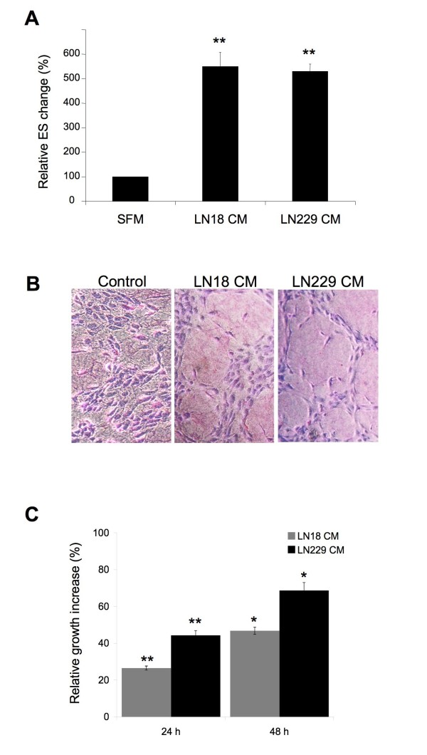
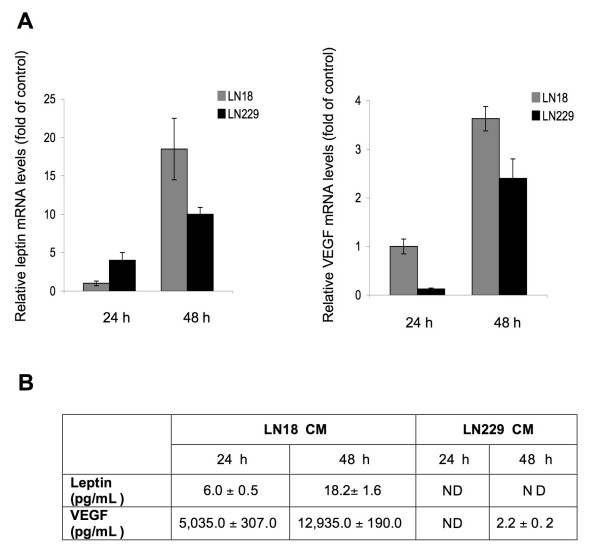
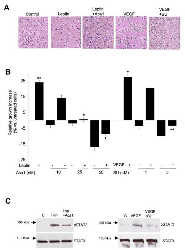
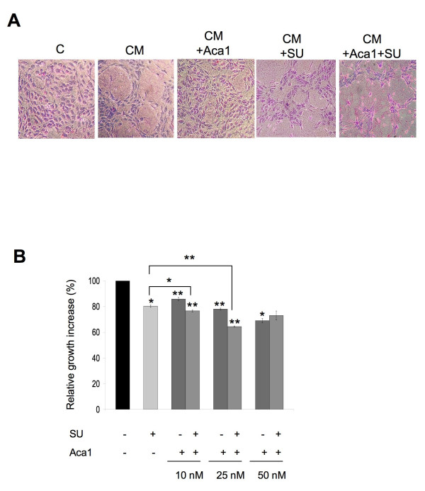
Results: We found that GBM cell lines LN18 and LN229 express leptin mRNA and LN18 cells secrete detectable amounts of leptin protein. Both lines also expressed and secreted VEGF. The conditioned medium (CM) of LN18 and LN 229 cultures as well as 200 ng/mL pure leptin or 50 ng/mL pure VEGF stimulated proliferation of human umbilical vein endothelial cells (HUVEC) at 24 h of treatment. Mitogenic effects of CM were ~2-fold greater than that of pure growth factors. Furthermore, CM treatment of HUVEC for 24 h increased tube formation by ~5.5-fold, while leptin increased tube formation by ~ 80% and VEGF by ~60% at 8 h. The mitogenic and angiogenic effects of both CM were blocked by Aca 1, a peptide ObR antagonist, and by SU1498, which inhibits the VEGF receptor. The best anti-angiogenic and cytostatic effects of Aca1 were obtained with 10 nM and 25 nM, respectively, while for SU1498, the best growth and angiogenic inhibition was observed at 5 μM. The combination of 5 μM SU1498 and Aca1 at 25 nM (growth inhibition) or at 10 nM (reduction of tube formation) produced superior effects compared with single agent treatments.

Conclusions: Our data provide the first evidence that LN18 and LN 229 human GBM cells express leptin mRNA and might produce biologically active leptin, which can stimulate tube formation and enhance proliferation of endothelial cells. Furthermore, we demonstrate for the first time that a peptide ObR antagonist inhibits proangiogenic and growth effects of leptin on endothelial cells, and that the pharmacological potential of this compound might be combined with drugs targeting the VEGF pathway.


Figures

Figure 1
Figure 1
CM derived from GBM cell lines LN18 and LN229 enhances angiogenic and mitogenic capabilities of HUVEC. A) Tube-like formation assay was performed in SFM or LN18 and LN229-derived CM mixed (1:1) with VCBM for 24 h, as described in Materials & Methods. The graph shows relative % increase of ES formed by HUVEC with the number in SFM taken as = 100%. Columns, mean of at least three independent experiments done in triplicates; bars, SE. ** P < 0,001 (vs. control SFM: VCBM). B) Representative phase contrast pictures (magnification 10×) of HUVEC in vitro angiogenesis in response to LN18 or LN229 CM for 24 h. SFM:VCBM treatment was used as control. The cultures were evaluated and photographed as described in Materials and Methods. C) HUVEC were grown in SFM:VCBM or LN18 and LN229-derived CM mixed (1:1) with VCBM for 24 or 48 h. The graph represents relative growth increase with SFM:VCBM taken as reference (0%). Columns, mean of at least three independent experiments done in triplicates; bars, SE. * P < 0,05 and ** P < 0,001 (vs. control SFM:VCBM).
Figure 2
Figure 2
GBM cells express leptin and VEGF. A) The abundance of leptin and VEGF mRNA was studied with qRT-PCR as described in Materials and Methods. The graphs represent leptin and VEGF mRNA levels relative to LN18 levels in SFM (= 1) ± SD. B) Leptin and VEGF secreted protein levels were measured in LN18 and LN229 CM by ELISA, as described in Materials and Methods. The values represent pg/mL per 9 × 10⁶ LN18 cells and 6 × 10⁶ LN229 cells ± SD.
Figure 3
Figure 3
Leptin and VEGF enhance angiogenesis and growth of HUVEC. ObR and VEGFR inhibitors counteract these effects. A) Representative phase contrast images (magnification 10×) of ES formed by HUVEC treated with 100 ng/mL leptin and/or with Aca1 or with 50 ng/mL VEGF and/or with SU1498 or left untreated (control) for 8 h. B) HUVEC growing in VCBM were treated for 24 h with 200 ng/mL leptin and/or Aca1 10, 25 or 50 nM or with 50 ng/mL VEGF and/or or with SU1498 1 and 5 μM or left untreated (control). The graph represents relative growth increase with untreated cells taken as reference (0%). Columns, mean of at least three independent experiments done in triplicates; bars, SE. * P < 0,05 and ** P < 0,01 (Leptin or VEGF vs. untreated cells, leptin + Aca1 vs. leptin or VEGF + SU1498 vs. VEGF). C) HUVEC were pretreated for 1 h with ObR or VEGFR inhibitors and then treated with 200 ng/mL leptin or 50 ng/mL VEGF for 15 min or left untreated as described in Materials and Methods. Levels of pSTAT3 and total STAT3 (~88 kDa) were determined by WB in 50-70 μg of total cell lysates using specific Abs, as described in Material and Methods. Arrows indicate the position of 105 kDa molecular weight marker.
Figure 4
Figure 4
Aca1 and SU1498 inhibit angiogenic and mitogenic effects of LN18 CM. A) Representative phase contrast images (magnification 10×) of ES formed by HUVEC grown for 24 h in SFM or LN18-derived CM containing or not 50 nM Aca1 and/or 5 μM SU1498. B) HUVEC were grown for 48 h in CM mixed (1:1) with VCBM, containing 10, 25 or 50 nM Aca1 and/or 5 μM SU1498 as described in Materials and Methods. The graph represents relative growth increase, with CM taken as reference (100%). Columns, mean of at least three independent experiments done in triplicates; bars, SE. * P < 0,05 and ** P < 0,01 (CM + Aca1 or CM + SU1498 5 μM vs. CM alone, CM + Aca1/SU1498 5 μM vs. CM + Aca1 and CM + SU1498 5 μM).

Similar articles

Cited by

References

    1. Friedman JM, Halaas JL. Leptin and the regulation of body weight in mammals. Nature. 1998;395(6704):763–770. - PubMed
    1. Zhang F, Chen Y, Heiman M, Dimarchi R. Leptin: structure, function and biology. Vitam Horm. 2005;71:345–372. - PubMed
    1. Cao Y. Angiogenesis modulates adipogenesis and obesity. J Clin Invest. 2007;117(9):2362–2368. - PMC - PubMed
    1. Rahmouni K, Haynes WG. Endothelial effects of leptin: implications in health and diseases. Curr Diab Rep. 2005;5(4):260–266. - PubMed
    1. Vona-Davis L, Rose DP. Angiogenesis, adipokines and breast cancer. Cytokine Growth Factor Rev. 2009;20(3):193–201. - PubMed

Publication types

MeSH terms

LinkOut - more resources