. 2011 Jul 20:8:60.
doi: 10.1186/1742-4690-8-60.

Characterization of the HIV-1 RNA associated proteome identifies Matrin 3 as a nuclear cofactor of Rev function

Affiliations

Characterization of the HIV-1 RNA associated proteome identifies Matrin 3 as a nuclear cofactor of Rev function

Anna Kula et al. Retrovirology. .

Abstract

Background: Central to the fully competent replication cycle of the human immunodeficiency virus type 1 (HIV-1) is the nuclear export of unspliced and partially spliced RNAs mediated by the Rev posttranscriptional activator and the Rev response element (RRE).

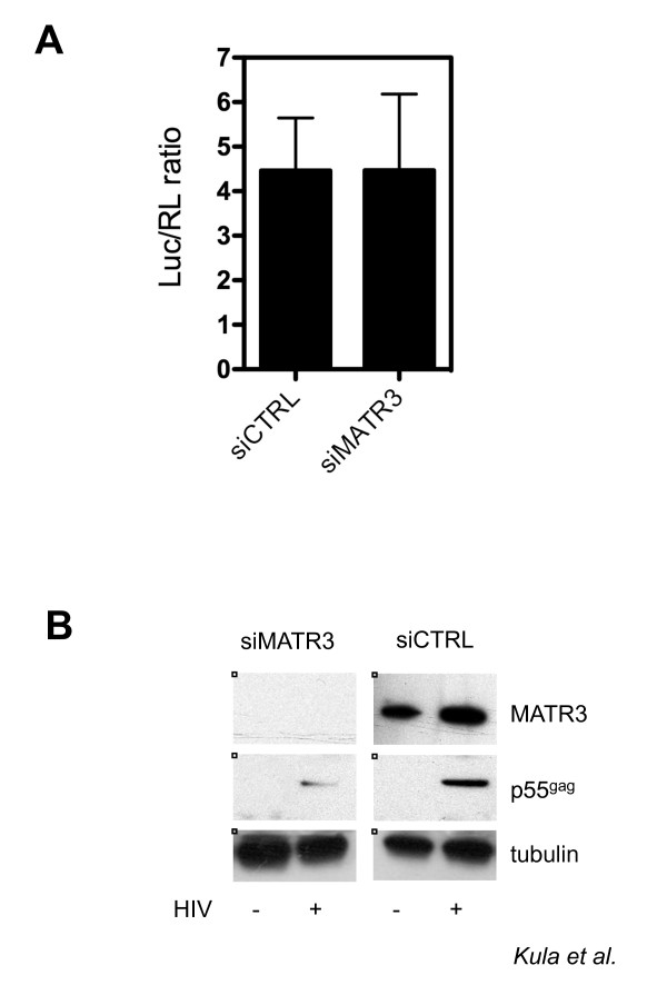
Results: Here, we introduce a novel method to explore the proteome associated with the nuclear HIV-1 RNAs. At the core of the method is the generation of cell lines harboring an integrated provirus carrying RNA binding sites for the MS2 bacteriophage protein. Flag-tagged MS2 is then used for affinity purification of the viral RNA. By this approach we found that the viral RNA is associated with the host nuclear matrix component MATR3 (Matrin 3) and that its modulation affected Rev activity. Knockdown of MATR3 suppressed Rev/RRE function in the export of unspliced HIV-1 RNAs. However, MATR3 was able to associate with Rev only through the presence of RRE-containing viral RNA.

Conclusions: In this work, we exploited a novel proteomic method to identify MATR3 as a cellular cofactor of Rev activity. MATR3 binds viral RNA and is required for the Rev/RRE mediated nuclear export of unspliced HIV-1 RNAs.


Figures

Figure 1
Figure 1
Detection and identification of HIV-1 RNA associated factors. A) Description of the HIV-1 constructs. Above an outline of the full-length viral genome, below the two constructs used in this work: HIVexo (carrying the MS2 binding sites after the SA7 splice site) and HIVintro (carrying the MS2 repeats in the intron). Black arrows indicate the RT-PCR primers listed in Table 2. The scheme is not drawn to scale. B) Pulldown of HIV-1 RNA and associated Tat. 293T cells expressing the indicated constructs were lysed and immunoprecipitated with anti-flag beads. Immunoblots with anti-GFP antibodies show Tat-CFP (lanes 1, 3, 5 7) and ECFPskl (lanes 5 and 7) expressed by the HIVintro construct. Tat could be immunoprecipitated only when the HIV-1 RNA is present and the association is disrupted by RNase treatment (compare lanes 6 and 8). IgH and IgL are the heavy and light chains of the immunoglobulins used in the immunoprecipitation. IP and WL stand for immunoprecipitation and whole cell's lysate, respectively. C) MS2-dependent pulldown of specific HIV-1 RNAs. U2OS clones and U2OS wt cells expressing Tat-CFP and flag-MS2nls were lysed and immunoprecipitated with anti-flag beads. RNA was extracted from immunoprecipitations and the RNA reverse-transcribed and PCR amplified with primers for β-actin mRNA (lanes 1-6), as well as with primers that differentiate spliced (lanes 7-12) and unspliced (lanes 13-18) forms of the HIV-1 RNAs which are outlined in Figure 1A.
Figure 2
Figure 2
Immunoprecipitation of HIV-1 RNA from nucleoplasmic fractions. A) Biochemical fractionation for the proteomic analysis. Nuclear extraction scheme showing the various phases of the protocol used to produce the nucleoplasmic fraction. B) Control of nuclear extraction in U2OS cells. The fractions obtained by the protocol outlined in Figure 2A were loaded on a gel for immunoblotting against α-tubulin (upper panel) that shows up only in the cytoplasmic fraction (CF) and against the nuclear protein RecQ (bottom panel) that was present only in the nucleoplasmic fraction (NF). C) Control of HIV-1 RNA associated factor Tat in the NF. Nuclear extracts from U2OS cells (mock), U2OS HIV_Exo_24 × MS2 (exo) or U2OS HIV_Intro_24 × MS2 (intro) were immunoprecipitated for HIV-1 RNA as described above, loaded on SDS-PAGE and blotted against GFP to detect the RNA-bound Tat-CFP protein (IP). Immunoblots for the nuclear extracts against GFP and flag-MS2nls (input) are shown. D) Pulldown of HIV-1 RNA and endogenous MATR3. Whole cell extracts from U2OS cells (mock), U2OS HIV_Exo_24 × MS2 (exo) or U2OS HIV_Intro_24 × MS2 (intro) were immunoprecipitated for HIV-1 RNA as described above, loaded on SDS-PAGE and blotted against MATR3 to detect the RNA-bound endogenous protein (IP). Immunoblots for the whole cell extracts against MATR3 and flag-MS2nls (input) are shown.
Figure 3
Figure 3
MATR3 is a post-transcriptional cofactor of HIV-1. A) MATR3 knockdown does not affect the luciferase activity. HeLa cells were transfected with the indicated siRNAs. After 48 hours siRNA-treated cells were transfected with the pNL4.3R-E-luc HIV-1 molecular clone and with pCMV-Renilla and harvested 24 hours later for luciferase assays. Relative Luc/RL expression was normalized to protein levels measured by Bradford assay. The results of three independent experiments are shown ± SD. B) MATR3 knockdown leads to decrease of the Gag expression from pNL4.3R-E-luc HIV-1 molecular clone. HeLa cells were transfected with the siRNA targeting MATR3 (siMATR3) or with a control siRNA (siCTRL). After 48 hours siRNA-treated cells were transfected with pNL4.3R-E-luc and harvested 24 hours later for immunobloting. Tubulin is the protein loading control.
Figure 4
Figure 4
MATR3 knockdown impairs Rev activity. A) Knockdown of MATR3 by siRNA. 293T cells were transfected either with siRNA targeting MATR3 (siMATR3) or with a control siRNA (siCTRL) and lysed after 72 hours for western blot analysis to assess the efficiency of MATR3 knockdown. Tubulin is the protein loading control. B) RT-PCR of spliced and unspliced HIV-1 RNA levels modulated by MATR3. Spliced (S) and unspliced (US) HIV-1 RNAs were detected (lanes 1-4, upper panel) simultaneously by RT-PCR on total RNA extracted from siRNA-treated 293T cells expressing vHY-IRES-TK, Tat and Rev-EGFP as indicated. RT-PCR amplification of an unrelated RNA was not affected (β-actin mRNA) (lanes 1-4, lower panel). Reactions without RT are shown to demonstrate lack of DNA contamination (lanes 5-8). Water (mock) was used as control of DNA contamination in the reaction. C) Quantitative analysis of unspliced HIV-1 RNA levels modulated by MATR3. Unspliced (US) viral RNA expression in siRNA treated 293T cells was assayed after transfection with vHY-IRES-TK, Tat and Rev-EGFP. Unspliced RNA levels were analyzed by quantitative real-time PCR and data normalized to β-mRNA expression. Data are presented as fold change, whereby siCTRL treated cells transfected with vHY-IRES-TK and Tat in the absence of Rev were set as 1. The results of three independent experiments are shown ± SD. The inhibition was significant (p = 0.00112). D) Rev-dependent expression of HIV-1 Gag (p17*). Western blot analysis of protein extracts from siRNA-treated 293T cells expressing vHY-IRES-TK, Tat and Rev-EGFP as indicated. p17* is the product of the truncated gag gene of the vHY-IRES-TK vector. Tubulin is the protein loading control. E) Quantitative analysis of unspliced HIV-1 RNA levels modulated by MATR3 in the nucleus and the cytoplasm. Unspliced (US) viral RNA expression in siRNA treated 293T cells was assayed after transfection with vHY-IRES-TK, Tat and Rev-EGFP. Unspliced RNA levels were analyzed by quantitative real-time PCR on nuclear (NF) and cytoplasmic fractions (CF). Data were normalized to β-mRNA expression and presented as fold changes, whereby siCTRL 293T treated cells transfected with vHY-IRES-TK and Tat and Rev-EGFP were set as 1. The results of three independent experiments are shown ± SD. The inhibition was significant (p = 0.00091). F) Quantitative analysis of spliced HIV-1 RNA levels modulated by MATR3 in the nucleus and the cytoplasm. The experiment was conducted for spliced (S) HIV-1 RNA as described above (Figure 4E).
Figure 5
Figure 5
MATR3 overexpression promotes Rev activity. A) Quantitative analysis of unspliced HIV-1 RNA levels modulated by transfected MATR3. Unspliced (US) viral RNA expression in 293T cells was assayed after transfection with Flag-MATR3, vHY-IRES-TK, Tat and Rev-EGFP. Unspliced RNA levels were analyzed by quantitative real-time PCR and data normalized to β-mRNA expression. Data are presented as fold change, whereby 293T cells transfected with vHY-IRES-TK and Tat in the absence of Rev were set as 1. The results of three independent experiments are shown ± SD. The increase was significant (p = 0.01931). B) Transfected MATR3 upregulates Rev-dependent Gag translation. Western blot analysis of protein extracts from 293T cells expressing Flag-MATR3, vHY-IRES-TK, Tat and Rev-EGFP. p17* is the product of the truncated gag gene of the vHY-IRES-TKvector. Tubulin is the protein loading control
Figure 6
Figure 6
MATR3 interaction with Rev requires HIV-1 RNA. A) Whole cell lysates from 293T cells expressing vHY-IRES-TK and Tat with or without Rev-EGFP were subjected to immunoprecipitation with anti-MATR3 antibodies or with anti-IgG (mock). The IP were subjected to nuclease treatment and the proteins were detected by immunoblotting. B) Whole cell lysates from 293T cells expressing either vHY-IRES-TK, or v653RSN or v653SN together with Tat and Rev-EGFP were subjected to immunoprecipitation with anti-MATR3 antibodies. Immunoblots from whole cell extracts are shown on the left (input). Endogenous β-actin was used as loading control. The immunoblot for p17* shows lack of Gag expression for the RRE deficient v653SN construct (bottom panel). Immunoprecipitations are shown on the right (IP).

Similar articles

Cited by

References

    1. Cullen BR. Viral RNAs: lessons from the enemy. Cell. 2009;136:592–597. doi: 10.1016/j.cell.2009.01.048. - DOI - PMC - PubMed
    1. Malim MH, Hauber J, Le SY, Maizel JV, Cullen BR. The HIV-1 rev trans-activator acts through a structured target sequence to activate nuclear export of unspliced viral mRNA. Nature. 1989;338:254–257. doi: 10.1038/338254a0. - DOI - PubMed
    1. Sodroski J, Goh WC, Rosen C, Dayton A, Terwilliger E, Haseltine W. A second post-transcriptional trans-activator gene required for HTLV-III replication. Nature. 1986;321:412–417. doi: 10.1038/321412a0. - DOI - PubMed
    1. McLaren M, Marsh K, Cochrane A. Modulating HIV-1 RNA processing and utilization. Front Biosci. 2008;13:5693–5707. - PubMed
    1. Chang DD, Sharp PA. Regulation by HIV Rev depends upon recognition of splice sites. Cell. 1989;59:789–795. doi: 10.1016/0092-8674(89)90602-8. - DOI - PubMed

Publication types

MeSH terms