. 2011 Sep 9;286(36):31337-46.
doi: 10.1074/jbc.M111.233353. Epub 2011 Jul 19.

Specific enzyme complex of beta-1,4-galactosyltransferase-II and glucuronyltransferase-P facilitates biosynthesis of N-linked human natural killer-1 (HNK-1) carbohydrate

Affiliations

Specific enzyme complex of beta-1,4-galactosyltransferase-II and glucuronyltransferase-P facilitates biosynthesis of N-linked human natural killer-1 (HNK-1) carbohydrate

Tetsuya Kouno et al. J Biol Chem. .

Abstract

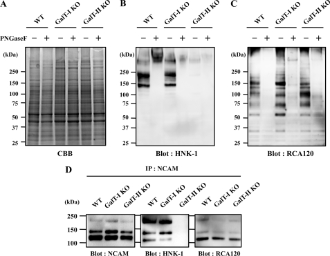
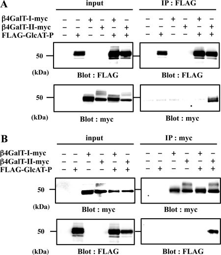
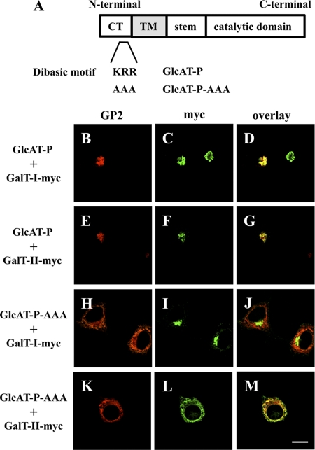
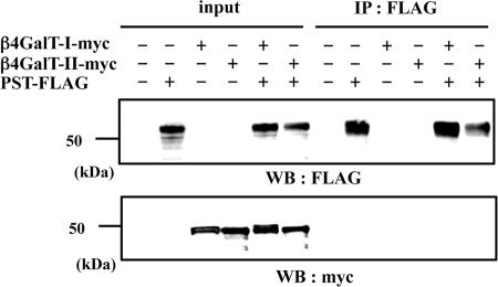
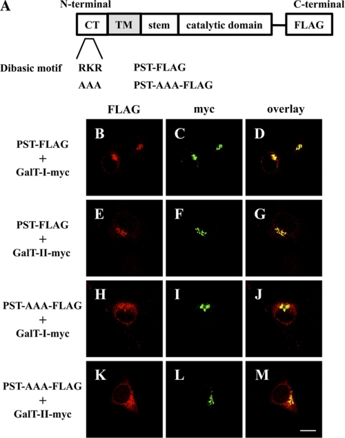
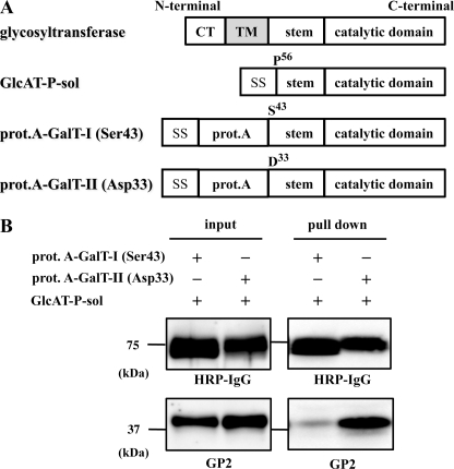
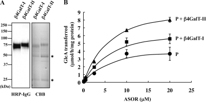
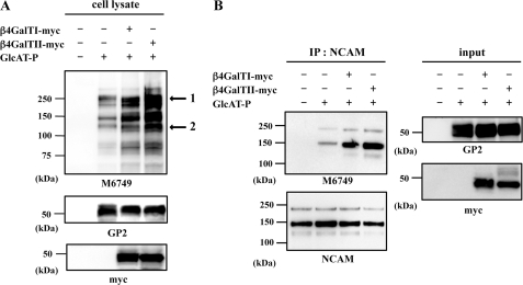
Human natural killer-1 (HNK-1) carbohydrate is highly expressed in the nervous system and is involved in synaptic plasticity and dendritic spine maturation. This unique carbohydrate, consisting of a sulfated trisaccharide (HSO(3)-3GlcAβ1-3Galβ1-4GlcNAc-), is biosynthesized by the successive actions of β-1,4-galactosyltransferase (β4GalT), glucuronyltransferase (GlcAT-P and GlcAT-S), and sulfotransferase (HNK-1ST). A previous study showed that mice lacking β4GalT-II, one of seven β4GalTs, exhibited a dramatic loss of HNK-1 expression in the brain, although β4GalT-I-deficient mice did not. Here, we investigated the underlying molecular mechanism of the regulation of HNK-1 expression. First, focusing on a major HNK-1 carrier, neural cell adhesion molecule, we found that reduced expression of an N-linked HNK-1 carbohydrate caused by a deficiency of β4GalT-II is not likely due to a general loss of the β1,4-galactose residue as an acceptor for GlcAT-P. Instead, we demonstrated by co-immunoprecipitation and endoplasmic reticulum-retention analyses using Neuro2a (N2a) cells that β4GalT-II physically and specifically associates with GlcAT-P. In addition, we revealed by pulldown assay that Golgi luminal domains of β4GalT-II and GlcAT-P are sufficient for the complex to form. With an in vitro assay system, we produced the evidence that the kinetic efficiency k(cat)/K(m) of GlcAT-P in the presence of β4GalT-II was increased about 2.5-fold compared with that in the absence of β4GalT-II. Finally, we showed that co-expression of β4GalT-II and GlcAT-P increased HNK-1 expression on various glycoproteins in N2a cells, including neural cell adhesion molecule. These results indicate that the specific enzyme complex of β4GalT-II with GlcAT-P plays an important role in the biosynthesis of HNK-1 carbohydrate.


Figures

FIGURE 1.
FIGURE 1.
Expression of HNK-1 carbohydrate and N-acetyllactosamine in mouse brains. Membrane fractions of 4-week-old C57BL/6 (WT), β4GalT-I-deficient (GalT-I-KO), and β4GalT-II-deficient (GalT-II-KO) mouse brain were subjected to SDS-PAGE followed by Coomassie Brilliant Blue (CBB) staining (A), Western blotting with HNK-1 mAb (B), or lectin blotting with RCA120 (C). D, membrane fractions of 4-week-old mouse brains were immunoprecipitated (IP) with anti-NCAM antibody, subjected to SDS-PAGE, and blotted with anti-NCAM mAb, HNK-1 mAb, and RCA120. PNGase F, peptide:N-glycosidase F.
FIGURE 2.
FIGURE 2.
Co-immunoprecipitation of GlcAT-P and β4GalT-II in N2a cells. Lysate of N2a cells transiently expressing FLAG-GlcAT-P, β4GalT-I-myc, or β4GalT-II-myc was immunoprecipitated (IP) with anti-FLAG pAb (A, IP: FLAG) or anti-Myc pAb (B, IP: myc), subjected to SDS-PAGE, and Western-blotted (Blot) with anti-FLAG mAb or anti-Myc mAb. To examine the level of each protein, the cell lysates were directly subjected to SDS-PAGE and Western blotting with anti-FLAG mAb or anti-Myc mAb (input).
FIGURE 3.
FIGURE 3.
ER retention assays using GlcAT-P-AAA. A, schematic diagrams of GlcAT-P and GlcAT-P-AAA. CT, cytoplasmic tail, TM, transmembrane domain. B–M, N2a cells were transiently co-transfected with GlcAT-P or GlcAT-P-AAA and β4GalT-I-myc or β4GalT-II-myc. GlcAT-P and GlcAT-P-AAA were detected with GP2 pAb (B, E, H, and K) and Alexa 546-conjugated secondary antibodies. β4GalT-myc (I and II) was detected with anti-Myc mAb (C, F, I, and L) and Alexa 488-conjugated secondary antibodies. D, G, J, and M, overlaid images. Bar, 10 μm.
FIGURE 4.
FIGURE 4.
Immunoprecipitation using PST-FLAG and β4GalT-myc in N2a cells. Lysate of N2a cells transiently expressing PST-FLAG, β4GalT-I-myc, or β4GalT-II-myc were immunoprecipitated (IP) with anti-FLAG pAb (IP: FLAG), subjected to SDS-PAGE, and Western-blotted with anti-FLAG mAb or anti-Myc mAb. To examine the level of each protein, the cell lysates were directly subjected to SDS-PAGE and then Western blotting (WB) with anti-FLAG mAb or anti-Myc mAb (input).
FIGURE 5.
FIGURE 5.
ER retention assays using PST-AAA-FLAG. A, schematic diagrams of PST-FLAG and PST-AAA-FLAG. B–M, N2a cells were transiently co-transfected with PST-FLAG or PST-AAA-FLAG and β4GalT-I-myc or β4GalT-II-myc. PST-FLAG or PST-AAA-FLAG was detected with anti-FLAG pAb (B, E, H, and K), and Alexa 546-conjugated secondary antibodies. β4GalT-myc (I and II) was detected with anti-Myc mAb (C, F, I, and L) and Alexa 488-conjugated secondary antibodies. D, G, J, and M, overlaid images. Bar, 10 μm. CT, cytoplasmic tail, TM, transmembrane domain.
FIGURE 6.
FIGURE 6.
Pulldown assays using soluble forms of GlcAT-P-sol, prot.A-GalT-I, and prot.A-GalT-II. A, schematic diagrams of GlcAT-P-sol, prot.A-GalT-I (Ser-43), and prot.A-GalT-II (Asp-33). SS, signal sequence. B, culture medium of N2a cells transiently expressing GlcAT-P-sol and prot.A-GalT-I (Ser-43) or prot.A-GalT-II (Asp-33) was incubated with IgG-Sepharose TM6 Fast Flow (pulldown), subjected to SDS-PAGE, and Western-blotted with HRP-conjugated normal rabbit IgG or GP2 pAb. To examine the level of each protein, the culture medium was directly subjected to SDS-PAGE and then Western-blotted with HRP-conjugated normal rabbit IgG or GP2 pAb (input). CT, cytoplasmic tail, TM, transmembrane domain.
FIGURE 7.
FIGURE 7.
Effect of the co-presence of β4GalT-II on the in vitro glucuronyltransferase reaction of GlcAT-P. A, prot.A-GalT-I (Ser-43) (β4GalT-I) or -II (Asp-33) (β4GalT-II) was expressed in COS-1 cells and purified from the culture media. The purified enzymes were subjected to Western blotting with HRP-conjugated rabbit IgG and Coomassie Brilliant Blue (CBB) staining. Asterisks indicate heavy and light chains of immunoglobulin derived from human IgG Sepharose. B, rate of glucuronyltransferase reaction of FLAG-P was measured in the absence of β4GalTs (P), or in the presence of prot.A-GalT-I (Ser-43) (P + β4GalT-I) or prot.A-GalT-II (Asp-33) (P + β4GalT-II) using different concentrations of ASOR as substrates. All experiments were employed in triplicate, and error bars indicate S.E.
FIGURE 8.
FIGURE 8.
Effect of the enzyme complex on HNK-1 expression in N2a cells. A, lysates of N2a cells transiently expressing GlcAT-P, β4GalT-I-myc, or β4GalT-II-myc were subjected to SDS-PAGE and then Western blotted with M6749 mAb, GP2 pAb, or anti-Myc mAb. Arrows indicate bands used for quantification by densitometric analyses. B, lysates of N2a cells transiently expressing GlcAT-P and β4GalT-I-myc or β4GalT-II-myc were immunoprecipitated (IP) with anti-NCAM mAb and subjected to SDS-PAGE and then Western-blotted with M6749 mAb or anti-NCAM mAb.

References

    1. Ohtsubo K., Marth J. D. (2006) Cell 126, 855–867 - PubMed
    1. Lowe J. B., Marth J. D. (2003) Annu. Rev. Biochem. 72, 643–691 - PubMed
    1. Sugimoto I., Futakawa S., Oka R., Ogawa K., Marth J. D., Miyoshi E., Taniguchi N., Hashimoto Y., Kitazume S. (2007) J. Biol. Chem. 282, 34896–34903 - PubMed
    1. Seko A. (2006) Trends Glycosci. Glycotechnol. 18, 209–230
    1. Seko A., Yamashita K. (2008) J. Biol. Chem. 283, 33094–33100 - PMC - PubMed

Publication types