. 2011 Sep 16;286(37):32661-71.
doi: 10.1074/jbc.M111.269399. Epub 2011 Jul 19.

Arrested oocyst maturation in Plasmodium parasites lacking type II NADH:ubiquinone dehydrogenase

Affiliations

Arrested oocyst maturation in Plasmodium parasites lacking type II NADH:ubiquinone dehydrogenase

Katja E Boysen et al. J Biol Chem. .

Abstract

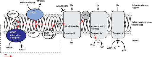
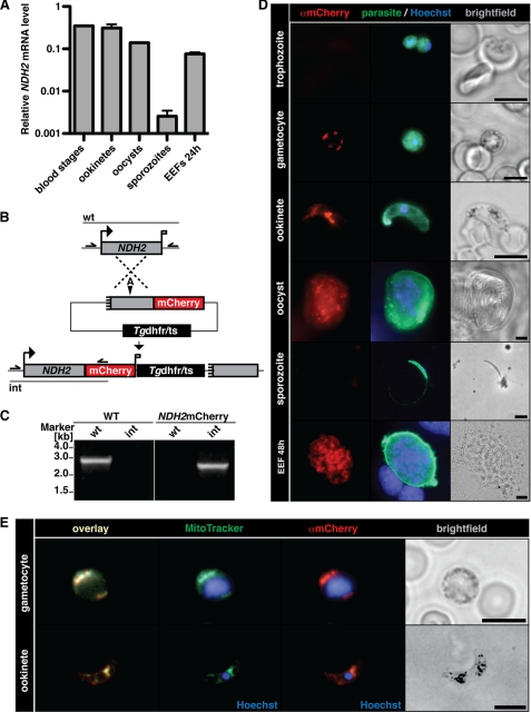
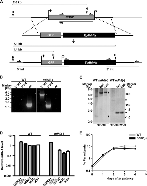
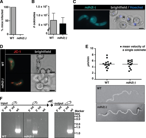
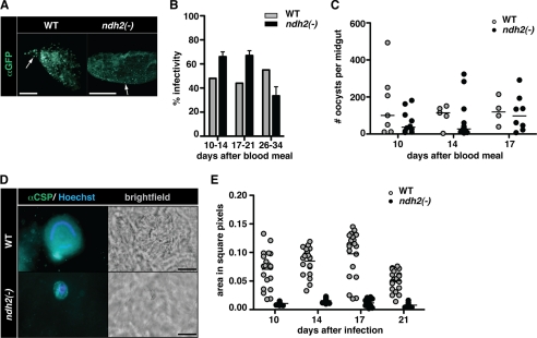
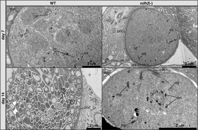
The Plasmodium mitochondrial electron transport chain has received considerable attention as a potential target for new antimalarial drugs. Atovaquone, a potent inhibitor of Plasmodium cytochrome bc(1), in combination with proguanil is recommended for chemoprophylaxis and treatment of malaria. The type II NADH:ubiquinone oxidoreductase (NDH2) is considered an attractive drug target, as its inhibition is thought to lead to the arrest of the mitochondrial electron transport chain and, as a consequence, pyrimidine biosynthesis, an essential pathway for the parasite. Using the rodent malaria parasite Plasmodium berghei as an in vivo infection model, we studied the role of NDH2 during Plasmodium life cycle progression. NDH2 can be deleted by targeted gene disruption and, thus, is dispensable for the pathogenic asexual blood stages, disproving the candidacy for an anti-malarial drug target. After transmission to the insect vector, NDH2-deficient ookinetes display an intact mitochondrial membrane potential. However, ndh2(-) parasites fail to develop into mature oocysts in the mosquito midgut. We propose that Plasmodium blood stage parasites rely on glycolysis as the main ATP generating process, whereas in the invertebrate vector, a glucose-deprived environment, the malaria parasite is dependent on an intact mitochondrial respiratory chain.


Figures

FIGURE 1.
FIGURE 1.
Hypothetical model of the mitochondrial NADH:oxidoreductase NDH2 in the mtETC of the malarial parasite. Shown is a schematic of the localization and biochemical function of PbNDH2 (dark blue ellipse). The precise localization to either the internal (mitochondrial matrix) or the external (inter-membrane space) side of the mitochondrial inner membrane remains to be determined. Together with four other Plasmodium mitochondrial dehydrogenases (DHOD) malate:quinone oxidoreductase (MQO), glycerol-3-phosphate dehydrogenase (G3PDH), and succinate dehydrogenase (SDH)), NDH2 likely acts as an electron donor in the mtETC, reducing ubiquinone (Q) to ubiquinol (QH2). DHOD (light blue ellipse), as part of the pyrimidine biosynthesis pathway, is thought to be essential. The antimalarial drug atovaquone targets the complex III, thereby inhibiting the reoxidation of QH2 to Q.
FIGURE 2.
FIGURE 2.
The type II NDH2 is a mitochondrial protein in the malaria parasite. A, expression profiling of NDH2 by quantitative real time PCR is shown. Data were normalized to GFP, which is constitutively expressed under the EF1α promoter. Note the abundant NDH2 transcripts in mixed blood stages and ookinetes and a prominent drop in mRNA levels in sporozoites. B, shown is the generation of NDH2-mCherry parasites. The NDH2 genomic locus was targeted with a replacement plasmid containing the C-terminal NDH2 fragment (gray box) fused in-frame to the mCherry coding sequence (red). In addition, the targeting plasmid contains the Tgdhfr/ts-positive selectable marker (black box). Upon a single crossover event, the targeting plasmid is expected to generate an mCherry-tagged NDH2 ORF and a non-expressed truncated fragment, shortened by 446 bp. Arrows and bars indicate specific primers and PCR fragments, respectively. A, AflII. C, genotyping of NDH2-mCherry parasites is shown. Integration of the mCherry tag (int) and absence of WT parasites (wt) confirmed the presence of a clonal NDH2-mCherry parasite population. D, epifluorescence images of NDH2-mCherry parasites during the entire life cycle are shown. Intra- or extracellular parasites were fixed, permeabilized, and stained with anti-mCherry (red) or anti-parasite (green) antibodies, and nuclei were visualized with Hoechst stain (blue). Bars, 5 μm. Note the absence of a mCherry signal in trophozoites and sporozoites. EEF, exo-erythrocytic form. E, NDH2 localizes to the mitochondrion of the parasite. Gametocytes and ookinetes of the NDH2-mCherry clone were incubated with MitoTracker Green FM (green), then fixed, permeabilized, and stained with anti-mCherry antibodies (red). Bars, 5 μm.
FIGURE 3.
FIGURE 3.
P. berghei NDH2 is dispensable for asexual blood stages. A, the wild-type NDH2 locus is targeted with a linearized replacement plasmid containing the 5′- and 3′-UTRs of PbNDH2, GFP, and the positive selection marker Tgdhfr/ts. After double crossover homologous recombination, the NDH2 open reading frame is substituted by GFP and the selection marker, resulting in the loss-of-function ndh2(−) allele. GFP is now expressed under the PbNDH2 promoter and, therefore, indicates PbNDH2 promoter activity. Replacement- and WT-specific test primer combinations, expected PCR fragments, and predicted sizes of restriction endonuclease fragments are shown as arrows, lines, and white bars, respectively. H, HindIII; N, NcoI. B, confirmation of the NDH2 gene disruption by replacement-specific PCR analysis with primer combinations that amplify a signal in the recombinant locus (5int and 3int) only. The absence of a WT-specific signal in the clonal ndh2(−) population confirms the purity of the mutant parasite line. C, a Southern blot confirms the desired NDH2 deletion in two clones from two independent transfections (ko1 and ko2). Fragments are marked with asterisks. Restriction sites used for the digest of genomic DNA are indicated at the bottom. The 5′ flank of the targeting vector was used as probe for the Southern blot. D, quantitative RT-PCR from WT and ndh2(−) mixed blood stages is shown. Shown are transcript levels for glycerol-3-phosphate dehydrogenase (G3PDH; gi:68071805), NDH2, dihydroxyorotate dehydrogenase (DHOD; gi:68074653), malate:quinone oxidoreductase (MQO; gi:68075787), and succinate dehydrogenase (SDH; gi:68063151). Data were normalized to the putative aspartyl-tRNA synthetase (PBANKA_021020). Note the depletion of NDH2 transcripts in ndh2(−) parasites. E, ndh2(−) parasites cause high level parasitemia in vivo. Displayed are in vivo growth curves of WT (gray) and knock-out parasites (black). Animals (n = 3) were injected intravenously with 1,000,000 asexual parasites of the respective parasite populations. Parasitemia was determined every 24 h after infection by microscopic examination of Giemsa-stained blood smears.
FIGURE 4.
FIGURE 4.
Ablation of NDH2 does not affect host switch from mammals to the insect vector. A, shown is a Plasmodium transmission experiment. Naïve mice were exposed to WT- or ndh2(−)-infected A. stephensi mosquitoes and examined daily for a blood stage infection. Data are from 3 separate experiments, with 2 ndh2(−) clones generated through separate transfection experiments and a total of 10 recipient mice. B, ookinete formation is not affected by the absence of NDH2. Ookinetes were formed in vitro in culture medium and quantified. Data are based on four in vitro cultures for each WT and ndh2(−). C, shown is epifluorescence live microscopy of a ndh2(−) ookinete. GFP expression confirms activity of the NDH2 promoter during sexual differentiation, ookinete viability, and successful integration of GFP into the NDH2 locus. Bar, 5 μm. D, mitochondrial membrane potential is detectable in WT and ndh2(−) ookinetes. Ookinetes were stained with the live stain JC-1, which forms red fluorescent aggregates in mitochondria if the membrane potential is intact. Bar, 5 μm. E, ookinete velocity is not affected by ablation of NDH2. Ookinetes in Matrigel were filmed for ∼10 min, and their tracks were quantified. Representative images of an ndh2(−) and a WT ookinete track (bottom) and mean velocity of WT ookinetes (n = 15) and ndh2(−) ookinetes (n = 14) (top) are shown. Note that all ndh2(−) ookinetes displayed gliding locomotion (n = 43).3 F, inability of ndh2(−) parasites to complete the life cycle is shown. A mix of both WT and KO ookinetes was membrane-fed to mosquitoes. After bite back, only WT blood stages could be detected in mice. Integration of KO construct and the absence of WT was monitored with PCR on genomic DNA derived from blood stage infection before setting up and mixing ookinete cultures (input) and after bite back (output).
FIGURE 5.
FIGURE 5.
ndh2(−) parasites are arrested in oocyst maturation. A, shown is successful colonization of midguts after transmission to A. stephensi. Mosquitoes were allowed to feed on WT- and ndh2(−)-infected mice, and midguts were removed 17 days later. Oocysts are visualized with an anti-GFP antibody. Note that ndh2(−) oocysts are much smaller. Arrows point at single oocysts. Bar, 500 μm. B, infectivity of WT- and ndh2(−)-infected A. stephensi midguts were isolated on the days indicated after a blood meal on infected mice and submitted to immunofluorescence analysis. The infectivity was similar in WT- and ndh2(−)-infected mosquitoes. Experiments were performed with two ndh2(−) clones generated through separate transfection experiments. C, oocyst numbers are similar in WT- and ndh2(−)-infected mosquitoes. Oocysts were scored from infected midguts between days 10 and 17 after the blood meal. The median is shown. The Mann-Whitney test was applied for every day indicated and revealed no significant differences in oocyst numbers. D, arrested oocyst growth of ndh2(−) parasites. Shown are representative oocysts from WT- and ndh2(−)-infected mosquitoes. Oocysts were stained with an anti-circumsporozoite protein (αCSP) antibody 16 days after infection. Note that ndh2(−) oocysts are clearly visible but significantly smaller than WT oocysts. WT oocysts show the typical circular stain of nuclei in sporoblasts. In ndh2(−) oocysts DNA can be stained but appears unorganized. Bar, 20 μm. E, shown is a quantification of oocyst size. Oocyst sizes in square pixels were measured using ImageJ. WT, n = 40; ndh2(−), n = 40.
FIGURE 6.
FIGURE 6.
Morphology of ndh2(−) and WT oocysts. Transmission electron microscopy shows low levels of organization in ndh2(−) oocysts and no sign of sporogony. Cristate mitochondria can be detected in both WT and KO oocysts. Note that ndh2(−) oocysts contain remnants of ookinete organelles, including the apical complex (lower right panel). Structures such as cristae and malaria pigment were identified by comparison with published electron microscope pictures (65, 66). apc, apical complex; apr, apical polar ring; cr, cristae; mi, mitochondrion; mn, micronemes; mp, malaria pigment; mt, microtubules; ooc, oocyst wall; sf, spindle fibers. sp, sporozoite.
FIGURE 7.
FIGURE 7.
NDH2 is essential for oocyst maturation only. A, shown is a schematic of WT (top) and ndh2(−) (bottom) cDNA isolated after the complementation experiment. Primers are indicated with arrows. Diagnostic fragments, i.e. ndh2(−) GFP for ndh2(−) parasites and Hsp70 as a positive control, are shown as lines. B, shown is the presence of ndh2(−) parasites in oocyst- and salivary gland-derived sporozoites after a blood meal on a mouse infected with a mixture of WT and ndh2(−) parasites (input in C). Real time-PCR products were analyzed by gel electrophoresis. ndh2(−)GFP was amplified in all mixed samples (WT/ndh2(−)) but not in WT. C, ndh2(−) sporozoites establish a blood stage infection. After bite back and isolation of Plasmodium genomic DNA, ndh2(−) blood stage parasites could be detected (output) by diagnostic PCR. Primers correspond to those shown in Fig. 3A.

References

    1. World Health Organization (2010) World Malaria Report 2010, World Health Organization Press, Geneva, Switzerland
    1. World Health Organization (2010) Global Report on Antimalarial Drug Efficacy and Drug Resistance: 2000–2010, World Health Organization Press, Geneva, Switzerland
    1. Petersen I., Eastman R., Lanzer M. (2011) FEBS Lett. 585, 1551–1562 - PubMed
    1. Painter H. J., Morrisey J. M., Mather M. W., Vaidya A. B. (2007) Nature 446, 88–91 - PubMed
    1. Mather M. W., Vaidya A. B. (2008) J. Bioenerg. Biomembr. 40, 425–433 - PubMed

Publication types

MeSH terms

Substances

LinkOut - more resources