. 2011 Aug;138(16):3545-55.
doi: 10.1242/dev.068148. Epub 2011 Jul 19.

Notch and Ras promote sequential steps of excretory tube development in C. elegans

Affiliations

Notch and Ras promote sequential steps of excretory tube development in C. elegans

Ishmail Abdus-Saboor et al. Development. 2011 Aug.

Abstract

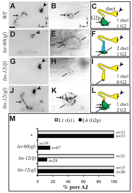
Receptor tyrosine kinases and Notch are crucial for tube formation and branching morphogenesis in many systems, but the specific cellular processes that require signaling are poorly understood. Here we describe sequential roles for Notch and Epidermal growth factor (EGF)-Ras-ERK signaling in the development of epithelial tube cells in the C. elegans excretory (renal-like) organ. This simple organ consists of three tandemly connected unicellular tubes: the excretory canal cell, duct and G1 pore. lin-12 and glp-1/Notch are required to generate the canal cell, which is a source of LIN-3/EGF ligand and physically attaches to the duct during de novo epithelialization and tubulogenesis. Canal cell asymmetry and let-60/Ras signaling influence which of two equivalent precursors will attach to the canal cell. Ras then specifies duct identity, inducing auto-fusion and a permanent epithelial character; the remaining precursor becomes the G1 pore, which eventually loses epithelial character and withdraws from the organ to become a neuroblast. Ras continues to promote subsequent aspects of duct morphogenesis and differentiation, and acts primarily through Raf-ERK and the transcriptional effectors LIN-1/Ets and EOR-1. These results reveal multiple genetically separable roles for Ras signaling in tube development, as well as similarities to Ras-mediated control of branching morphogenesis in more complex organs, including the mammalian kidney. The relative simplicity of the excretory system makes it an attractive model for addressing basic questions about how cells gain or lose epithelial character and organize into tubular networks.


Figures

Fig. 1.
Fig. 1.
Timeline of excretory system development. (A) Schematics of excretory canal cell (red, ABplpappaap), duct (yellow, ABplpaaaapa), G1 (blue, ABprpaaaapa), G2 (green, ABplapaapa) and W (green, ABprapaapa) at different developmental stages, based on Sulston et al. (Sulston et al., 1983), prior electron microscopy (Stone et al., 2009) and this work. Dark black lines, apical junctions; dotted line, duct auto-fusion; arrow, pore autocellular junction; arrowhead, duct-canal cell intercellular junction; bracket, duct cell body. Not shown are the non-essential excretory gland cells, which also connect to the duct-canal junction (Nelson et al., 1983; Nelson and Riddle, 1984). (B-E) Progressively older ventral enclosure stage embryos. (B-C,E) Ventral views. GFP::MLS-2 marks the presumptive duct and G1 pore nuclei. DLG-1::GFP marks epidermal cell junctions in B and E, which are confocal projections. (B) The presumptive duct and G1 initially lack junctions. (C) The presumptive duct is closer to the canal cell than is the presumptive G1. (D) TEM of a wild-type embryo at a similar stage to C, with cells pseudo-colored as in A. Transverse anterior view. The presumptive duct and G1 have met at the ventral midline. The duct makes extensive contact with the canal cell, while G1 is excluded. No epithelial junctions or lumen are detectable. (E) G1 moves ventrally. The asterisk indicates the site of future G1 pore opening between G2 and W epidermal cells (see also Fig. S1 in the supplementary material). (F-L) Left lateral views. (F) 1.5-fold stage embryo immunostained for DLG-1, showing newly formed autocellular junctions (inset) just before duct auto-fusion. (G-K) L1 larvae. The box in G′ indicates the region magnified in H. AJM-1::GFP marks junctions. The duct no longer has an autocellular junction. (I) lin-48p::mcherry marks the duct. (J) dct-5p::mcherry marks the duct and G1 pore in early L1 and (K) the duct and G1 in late L1 after G1 withdrawal and G2 entry. (L) Adult canal cell marked with vha-1p::GFP. Note that the canal cell elongates extensively.
Fig. 2.
Fig. 2.
let-60/Ras promotes the duct versus G1 pore fate. (A-J′) AJM-1::GFP (left column) and lin-48p::GFP or (C′) dct-5p::mCherry (middle column) expression in L1 larvae of the indicated genotypes. Lateral views, with schematic interpretations (right column) and symbols as in Fig. 1. Colors represent lineal identity, not fate. Mutants with reduced signaling usually have two pore-like cells with autocellular junctions, but as fluid (carat) accumulates during L1 (C), large junctional rings (asterisk) are common. In (C′), white arrows indicate two stacked pore-like cells. Mutants with increased signaling have a seamless binucleate duct that connects to the ventral epidermis. (K,L) Quantification of marker phenotypes. Note that some mutants with `0 G1' have defects in cell stacking and tubulogenesis rather than in cell fate specification (see Fig. 4). Scale bar: 2 μm.
Fig. 3.
Fig. 3.
lin-3/EGF, let-23/EGFR and lin-12/Notch reporter expression in the excretory system. (A,C) LET-23::GFP is expressed in the presumptive duct and G1 pore at ventral enclosure (A) and in the duct (bracket) at 3-fold (C). (B,D) lin-3p::GFP is expressed in the canal cell from ventral enclosure (B) through L1 (D). (E) LIN-12::GFP is expressed in the presumptive G2 and W but not in the presumptive duct or pore at ventral enclosure. In A, C and E, embryos were co-stained with anti-GFP and either anti-MLS-2 or anti-DLG-1 to mark the duct and G1 pore (n>10 each). Scale bars: 5 μm.
Fig. 4.
Fig. 4.
let-60/Ras hypomorphs reveal defects in cell competition and stacking. (A,B,E,F) Wild-type. (C,D,G-J) let-60(n2021rf). (A,C) AJM-1::GFP in threefold embryos, ventral view. Lines indicate ventral junctions between the presumptive G1 or duct and the epidermis. (E,G,I,K) AJM-1::GFP and dct-5p::mCherry in early L1s, lateral view. Asterisks indicate ventral cells with neither duct-like nor pore-like morphology. In let-60(n2021rf) mutants, the presumptive duct and G1 adopt adjacent positions in the epidermis (C,D) and one cell usually reaches from the ventral epidermis to the canal cell (G-J). The lineal identity of this cell is unknown and may be variable. (K) Quantification of adjacent defects in early L1 larvae from heterozygous versus homozygous mutant mothers. Animals with no G1 pore autocellular junction or with a single autocellular junction that stretched from the ventral epidermis to the canal junction were scored as `adjacent'.
Fig. 5.
Fig. 5.
The canal cell is required for duct and G1 pore stacking and tubulogenesis. (A-I,L) AJM-1::GFP. D,G,H also contain lin-48p::GFP. (J,K) Schematic diagrams. (A-C) Early threefold embryos, ventral view. (D-F) Early L1s, ventral view. (G-L) Early L1s, lateral view. In wild-type (A), the G1 pore contacts G2 and W in the ventral epidermis. In lag-1(RNAi) (B) or lin-12(n941) glp-1(q46) double mutants (C,F,I), the presumptive duct and G1 pore (lines) both contact the epidermis and lack autocellular junctions. In lag-2(q420rf) mutants (D,E,G,H), the presence of a canal cell (D,G) correlates with normal duct and G1 pore morphology. aff-1(tm2214) (E) has no impact on the lag-2(q420rf) phenotype. (L) Ablation of the canal cell mother eliminates the G1 pore autocellular junction. (M) Quantification of junction phenotypes in early L1 larvae. Animals with no G1 pore autocellular junction were scored as `adjacent'. Scale bar: 2 μm.
Fig. 6.
Fig. 6.
sos-1 temperature shift experiments reveal continued requirements during duct morphogenesis and differentiation. (A) sos-1(cs41ts) lethality at 25°C is rescued by let-60(n1046gf) or lin-1(e1275lf). n>50 for each genotype. (B,C) sos-1(ts) animals bearing AJM-1::GFP or lin-48p::GFP markers were upshifted or downshifted at the stages indicated. n≥20 for each time point. sos-1 is required before the 1.5-fold stage to promote lin-48p::GFP duct marker expression (B) or duct auto-fusion (C). (D) The sos-1(ts) temperature-sensitive period (TSP) for lethality extends from the bean stage of embryogenesis to L2. The majority of animals upshifted before L2 arrested with excretory abnormalities (see C,E,F). Animals upshifted during L2 displayed a scrawny phenotype similar to that reported for egl-15/FGFR mutants (DeVore et al., 1995; Roubin et al., 1999). (E-F′) Fluid (carats) accumulated in or near the duct in threefold upshifted sos-1(ts) animals (E',F'), while AJM-1::GFP (E) and lin-48p::GFP (F) patterns were unaffected in these same animals. Scale bar: 2 μm.
Fig. 7.
Fig. 7.
G1 withdrawal and G2 entry can occur independently. AJM-1::GFP in L4 larvae. (A,D,G,J) lateral views. (B,E,H,K) ventral views. (C,F,I,L) Schematic diagrams. (A-C) In wild type, G2p forms the pore. (D-F) In let-60(n1046gf) mutants, G2p usually wraps around the base of the duct. (G-I) In lin-12(n941lf) mutants, the duct attaches directly to the ventral epidermis after G1 withdrawal. (J-L) In lin-12(n137gf) mutants, the extra G2p cell wraps around the ventral base of the pore. Lines indicate ventral junctions with the epidermis. (M) Quantification of junction phenotypes. Scale bar: 2 μm.

Similar articles

Cited by

References

    1. Bao Z., Murray J. I., Boyle T., Ooi S. L., Sandel M. J., Waterston R. H. (2006). Automated cell lineage tracing in Caenorhabditis elegans. Proc. Natl. Acad. Sci. USA 103, 2707-2712 - PMC - PubMed
    1. Bar T., Guldner F. H., Wolff J. R. (1984). `Seamless' endothelial cells of blood capillaries. Cell Tissue Res. 235, 99-106 - PubMed
    1. Beitel G. J., Tuck S., Greenwald I., Horvitz H. R. (1995). The Caenorhabditis elegans gene lin-1 encodes an ETS-domain protein and defines a branch of the vulval induction pathway. Genes Dev. 9, 3149-3162 - PubMed
    1. Berry K. L., Bulow H. E., Hall D. H., Hobert O. (2003). A C. elegans CLIC-like protein required for intracellular tube formation and maintenance. Science 302, 2134-2137 - PubMed
    1. Bossinger O., Klebes A., Segbert C., Theres C., Knust E. (2001). Zonula adherens formation in Caenorhabditis elegans requires dlg-1, the homologue of the Drosophila gene discs large. Dev. Biol. 230, 29-42 - PubMed

Publication types

MeSH terms